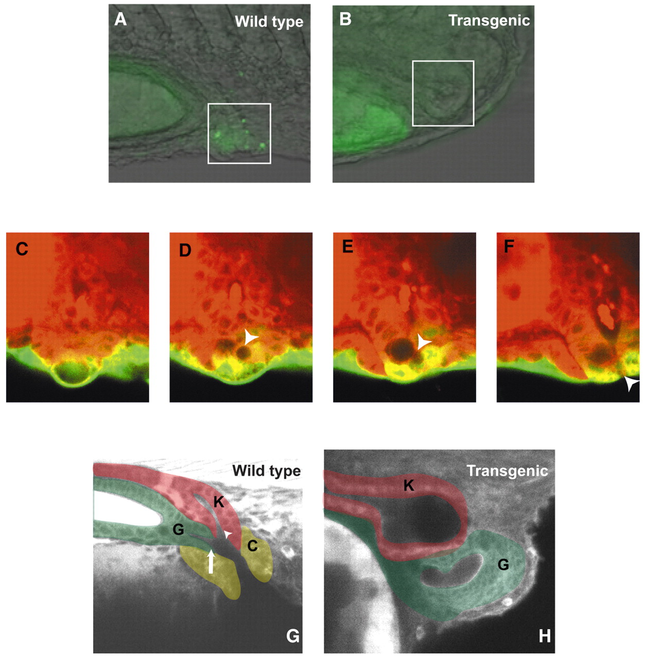
Fig. 6 Visualizing cloaca development in living embryos. (A,B) Acridine Orange staining in wild-type and transgenic embryos to assay cell death in the developing cloaca region. The posterior kidney region is boxed. (C-F) Detailed time lapse of an msxb-gfp embryo during opening of the presumptive cloaca. Note initially the kidney terminus, cup-shaped proctodeum, and epidermis (green; C) at 24-somites. A single vacuolated proctodeal cell (arrowhead in D-F) emerges and migrates to the ventral limit of the epidermis, where it forms a pore (F). At that point, the kidney terminus has connected to the epidermis and there is a continuous opening to the outside of the embryo (see also Movie 1 in the supplementary material for the full time lapse). (G,H) Excretory region of a wild-type and transgenic sibling larva, respectively, at 4 dpf. Regions of the excretory system have been pseudo-colored for identification: K, kidney (red); G, gut (green); C, cloaca (yellow). Arrowhead in G marks the kidney (urogenital) opening, and arrow marks the gut opening.
Image
Figure Caption
Figure Data
Acknowledgments
This image is the copyrighted work of the attributed author or publisher, and
ZFIN has permission only to display this image to its users.
Additional permissions should be obtained from the applicable author or publisher of the image.
Full text @ Development

