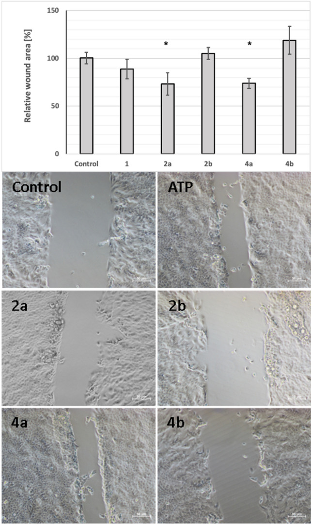
Figure 5
Migration of human keratinocytes after treatment with α-thio-modified ATP analogues. The rate of wound healing after 24 h treatment of HaCaT cells with 100 μM of α-thio-modified ATP derivatives. Upper panel: relative migration rate of HaCaT cells quantified based on the size of the uncovered area after 24 h incubation with tested compounds compared to the untreated cells (taken as control sample 100%). Data represent the means ± SEM from at least 3 independent experiments. Lower panel: the microscopy images of the wound area after 24 h incubation with tested compounds. *p < 0.05 compared to the control, Scale bars 50 μm. The initial sizes of the scratches are available in Supplementary Materials (Fig.

