IMAGE

Fig 4

ID
ZDB-IMAGE-240524-124
Source
Figures for Li et al., 2024
Image
Figure Caption

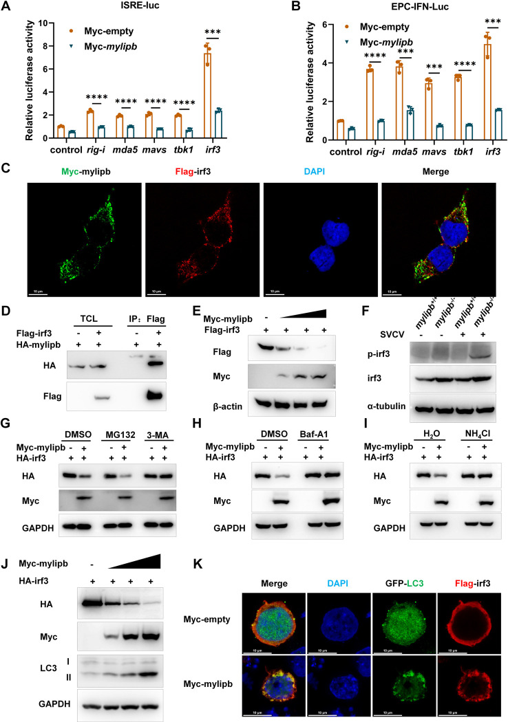
Fig 4 Mylipb interacts with irf3 and induces autophagic degradation of irf3.

(A) Overexpression of mylipb suppressed rig-i–, mda5- mavs-, tbk1-, and irf3-induced activation of ISRE luciferase activity in EPC cells. (B) Overexpression of mylipb suppressed rig-i–, mda5- mavs-, tbk1-, and irf3-induced activation of EPC–IFN promoter luciferase reporter (EPC-IFN-luc) activity in EPC cells. (C) Immunofluorescence staining showed the predominant colocalization of mylipb and irf3 protein in the cytoplasm. HEK293T cells were co-transfected with Myc-mylipb and Flag-irf3. After 24 h, the cells were fixed and observed by confocal microscopy. Red signals represent overexpressed irf3, green signals represent overexpressed mylipb. (D) Mylipb associated with irf3. HEK293T cells seeded in 100-mm dishes were transfected with the indicated plasmids (4 μg each). After 24 h, total cell lysates were immunoprecipitated (IP) with anti-Flag antibody conjugated agarose beads. Then, the immunoprecipitates and cell lysates were detected with anti-HA or anti-Flag Ab, respectively. (E) Mylipb induced the degradation of irf3 in a dose-dependent manner. HEK293T cells were transfected with Myc-empty, Myc-mylipb, and Flag-irf3 for 24 h, and then the cells were harvested to perform immunoblotting. (F) The protein level of irf3 and p-irf3 was higher in mylipb-null zebrfish liver compared with that in WT zebrafish. mylipb-null zebrafish (3 mpf) and their WT siblings (3 mpf) were i.p. injected with (+) or without (-) SVCV at 10 μl/individual for 48 h, then their livers were dissected for Western blot analysis using anti-irf3, anti-p-irf3. (G) Immunoblotting of lysates from HEK293T cells transiently transfected with Myc-empty, Myc-mylipb, and HA-irf3 for 24 h and then cultured in the presence of the proteasome inhibitor, MG132 (20 μM), autophagy inhibitor, 3-MA (1 mM),or DMSO for 12 h. (H) Immunoblotting of lysates from HEK293T cells transiently transfected with Myc-empty, Myc-mylipb, and HA-irf3 for 24 h and then cultured in the presence of the autophagy inhibitor, Baf-A1 (100 nM), or DMSO for 12 h. (I) Immunoblotting of lysates from HEK293T cells transiently transfected with Myc-empty, Myc-mylipb, and HA-irf3 for 24 h and then cultured in the presence of the lysosome inhibitor, NH4Cl (5 mM), or H2O for 12 h. (J) Immunoblotting of lysates from HEK293T cells transiently transfected with Myc-empty, Myc-mylipb, and HA-irf3 for 24 h. (K)) HEK293T cells were co-transfected with Myc-mylipb or pCMV-Myc plus Flag-irf3 and GFP-LC3. After 24 h, the cells were fixed and observed by confocal microscopy. Red signals represent overexpressed irf3, green signals represent LC3 positive autophagosome accumulation.

Acknowledgments
This image is the copyrighted work of the attributed author or publisher, and ZFIN has permission only to display this image to its users. Additional permissions should be obtained from the applicable author or publisher of the image. Full text @ PLoS Pathog.