
|
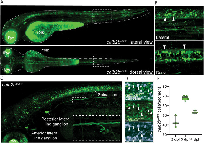
Fig. 1 Overall characterization of calb2beGFP transgenic line. A) Dorsal and lateral light-sheet images of the calb2beGFP transgenic line at 3 days post fertilization (dpf). B) Dorsal and lateral view of the spinal cord at 3 dpf. calb2beGFP positive primary and secondary motor neurons labelled by are indicated by white arrowheads. C) Confocal image with details of the lateral line from head to spinal cord at 3 dpf. D) Whole mount immunohistochemistry against calretinin in calb2beGFP larvae at 3 dpf. E) Quantification of eGFP-positive cells in calb2beGFP larvae from 2 to 4 dpf. Scale bar, 100 μm in A and B; 50 μm in C and D.
Reprinted from Developmental Biology, 508, Iglesias Gonzalez, A.B., Koning, H.K., Tuz-Sasik, M.U., van Osselen, I., Manuel, R., Boije, H., Perturbed development of calb2b expressing dI6 interneurons and motor neurons underlies locomotor defects observed in calretinin knock-down zebrafish larvae, 778777-87, Copyright (2024) with permission from Elsevier. Full text @ Dev. Biol.