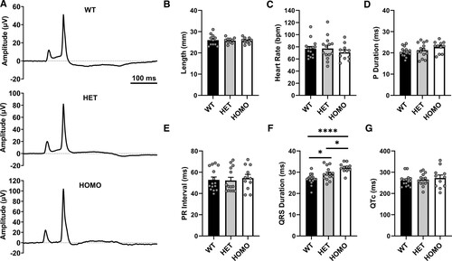
Fig. 2 mapre2 loss of function leads to conduction slowing in adult fish. Two-lead surface electrocardiogram (ECG) was performed in anesthetized fish from the mapre2 knockout (KO) line. A, Representative averaged ECG tracings demonstrating similarity to human ECG with the notable exception of the inverted T wave. B through G, In homozygous (HOMO) mapre2 KO, there is a nonsignificant increase in P-wave duration and a significant increase in QRS duration, suggesting ventricular conduction slowing, with heterozygotes showing an intermediate phenotype (1-way ANOVA, P<0.0001 followed by Tukey multiple comparisons test, P=0.0435 wild-type [WT] vs heterozygous [HET]; P=0.0314 HET vs HOMO; P<0.0001 WT vs HOMO; F). Each dot represents 1 fish.
Image
Figure Caption
Acknowledgments
This image is the copyrighted work of the attributed author or publisher, and
ZFIN has permission only to display this image to its users.
Additional permissions should be obtained from the applicable author or publisher of the image.
Full text @ Circ. Res.

