
Image
|
Figure Caption
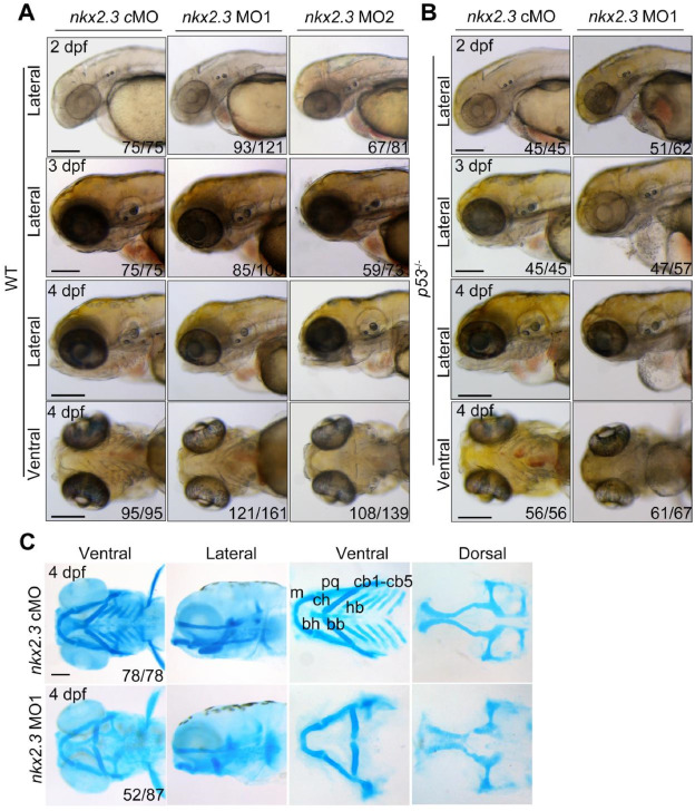
Fig. 1 Malformation of pharyngeal cartilages in nkx2.3 morphants. (A) Morphology of WT embryos injected with nkx2.3 MOs at indicated stages. Scale bars, 200 μm. (B) Morphology of p53 mutant embryos injected with nkx2.3 MOs at 4 dpf. Scale bars, 200 μm. (C) Detection of craniofacial cartilages formation using Alcian blue staining. The cartilages were shown anterior to the left and included the ceratobranchial (cb), ceratohyal (ch), Meckel's cartilage (m), basibranchial (bb), basihyal (bh), hypobranchial(hb), and palatoquadrate (pq) cartilages. Scale bars, 100 μm.
Figure Data
Acknowledgments
This image is the copyrighted work of the attributed author or publisher, and
ZFIN has permission only to display this image to its users.
Additional permissions should be obtained from the applicable author or publisher of the image.
Full text @ Heliyon