IMAGE

Figure 4.

ID
ZDB-IMAGE-240213-4
Source
Figures for Kahsay et al., 2024
Image
Figure Caption

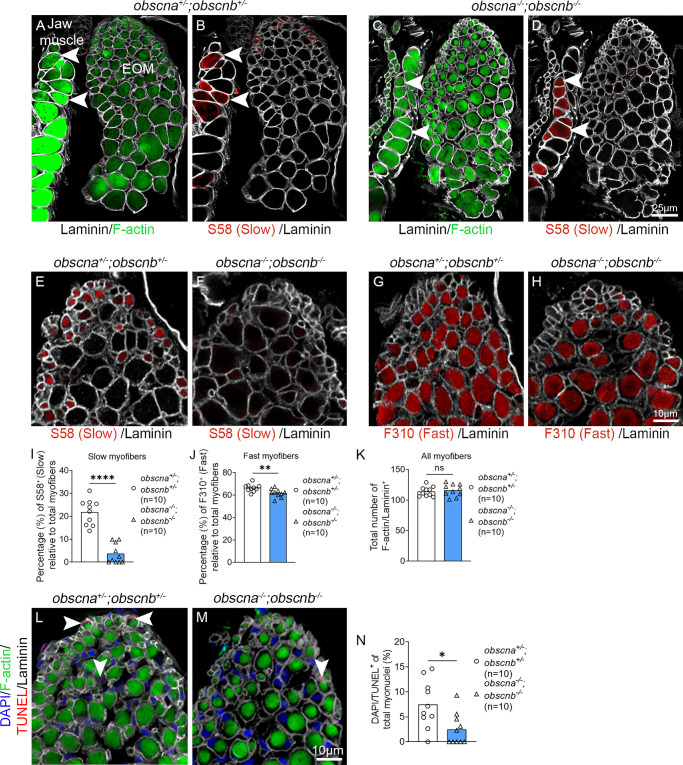
Figure 4.

Quantification of slow and fast myofibers in the EOMs of adult obscurin mutants and sibling controls. Cross-sections of 10-month-old adult zebrafish EOMs immunolabeled with phalloidin to identify all myofibers (labeling of F-actin by phalloidin, green in A and C), S58 to identify all myofibers containing slow MyHC (red in B, D–F), F310 to identify all myofibers containing fast MyHC (red in G and H). The contours of the myofibers were labeled by the antibody against laminin (gray in A–H). Arrowheads indicate examples of positively-labeled myofibers with phalloidin, which labels F-actin (in A and C) and slow MyHC S58 (in B and D) in closely located masticatory myofibers (left) in the same section. Quantification of slow myofibers (S58 positive in E and F and quantified in I), fast myofibers (F310 positive in G and H and quantified in J), and the total number of myofibers (K) in obscna/;obscnb/ and obscna+/;obscnb+/. (L, M) Cross-sections of obscna+/;obscnb+/ and obscna/;obscnb/ zebrafish EOMs immunolabeled with phalloidin, which labels F-actin, laminin, DAPI and TUNEL (arrowheads, red in L and M) to identify apoptotic myofibers. (N) The proportion of TUNEL-positive myofibers. Data in graphs are presented as mean ± SEM. *P < 0.05; **P < 0.01; ****P < 0.0001.

Acknowledgments
This image is the copyrighted work of the attributed author or publisher, and ZFIN has permission only to display this image to its users. Additional permissions should be obtained from the applicable author or publisher of the image. Full text @ Invest. Ophthalmol. Vis. Sci.