
|
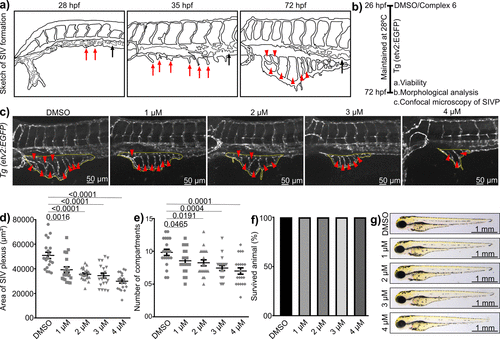
Fig. 9 Complex 6 inhibits blood vessel formation in a dose-dependent manner. (a) Graphic representation of subintestinal venous plexus (SIVP) formation in a wild-type embryo. Red arrows indicate angiogenic sprouts from the posterior cardinal vein (PCV, black arrow), around 28 and 48 hpf (hours post fertilization). Red arrowheads indicate vascular compartments in SIVPs at 72 hpf. (b) Schematic representation of the experimental procedures. (c) Maximum intensity confocal images of the SIVP region at 72 hpf transgenic embryos Tg(etv2:EGFP), expressing EGFP in the endothelial cells of the blood vessels. The SIVP area is shown with yellow dotted lines, and red arrowheads represent the compartments. (d) Dot plot depicting the area covered by SIVP (n = 20 from each treatment condition). (e) Quantification of the number of compartments in SIVPs (n = 20 from each treatment condition). (f) Embryo viability analysis. (g) The bright-field images of the embryos at 3 dpf (dpf = days post fertilization, scale: 1 mm). Figure S32 is the extended image. In panels (d) and (e), data are mean ± standard error of the mean (SEM), and each sample represents one animal.