IMAGE

Fig 5

ID
ZDB-IMAGE-240122-6
Source
Figures for Schumacher et al., 2024
Image
Figure Caption

Fig 5 
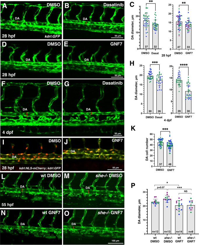
Inhibitors of Abl signaling reduce DA diameter in wild-type and she mutant embryos.

(A-E) Dorsal aorta diameter at 28 hpf is reduced in kdrl:GFP embryos treated with 5 μM Dasatinib or 1 μM GNF-7 compared to controls treated with 0.1% DMSO. (F-H) Embryos treated with 5 μM Dasatinib (F-H, left) or 1 μM GNF-7 (H, right) exhibit narrower DA at 4 dpf compared to controls treated with 1% DMSO (GNF-7) or 2% DMSO (Dasatinib treatments). (I-K) The number of cells in the DA is reduced in embryos at 28 hpf treated with 1 μM GNF-7 compared to control embryos treated with 0.1% DMSO. (L-O) GNF-7 treatment reverses DA enlargement in she mutant embryos. she+/-; kdrl:GFP adults were crossed to obtain she mutant embryos. Embryos were treated starting at 6 hpf with either 0.5 μM GNF-7 or 0.1% DMSO. Embryos were imaged at approximately 55 hpf and subsequently genotyped. DA measurements were performed blinded. Mid-trunk region is shown, anterior is to the left. Note the slightly wider DA (red line) in she-/- mutant embryos compared to wild-type (she+/+) siblings. DA is reduced in both wild-type and she mutant embryos treated with GNF-7. (P) Quantification of DA diameter in wild-type or she mutant embryos treated with GNF-7 or DMSO. In all graphs mean±SD is shown. Data points (shown in different colors) are combined from 2 (left graph C,H,K) or 3 (right graph C,P) independent experiments. Total number of embryos analyzed is shown at the bottom of each bar. *p<0.05, **p<0.01, ***p<0.001, ****p<0.0001, NS–not significant, Student’s t-test (C,H,K), or one-way ANOVA test, followed by multiple comparisons Fisher’s LSD (P).

Acknowledgments
This image is the copyrighted work of the attributed author or publisher, and ZFIN has permission only to display this image to its users. Additional permissions should be obtained from the applicable author or publisher of the image. Full text @ PLoS Genet.