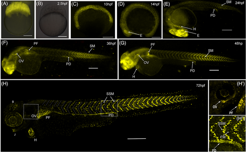
Fig. 4 mCherry expression begins just before somite formation. (A, B) mCherry fluorescence (shown in yellow) in 2 hpf zebrafish zygotes obtained from a wild-type fish crossed with a transgenic female (panel A) or transgenic male (panel B). (C-H) zygotic mCherry expression in zebrafish embryos obtained from crosses of transgenic males and wild-type females starts at 10 hpf (panel C). By the 14 hpf (panel D), mCherry is observed in the eye (E) and mesodermal tissue including the somites. At later stages (panels E-H), mCherry expression is clearly observed in the trunk superficial skeletal muscle (SSM), heart (H), pectoral fin (PF), otic vesicle (OV), pronephric duct (PD), jaw (J), and non-identified cells from the eye and brain (B). Inset H′ shows the reporter expression in the OV and PF. Inset H" shows mCherry in skeletal mononuclear muscle cells at the level of somites 14 to 16. Scale bars 200 μm.
Image
Figure Caption
Acknowledgments
This image is the copyrighted work of the attributed author or publisher, and
ZFIN has permission only to display this image to its users.
Additional permissions should be obtained from the applicable author or publisher of the image.
Full text @ Dev. Dyn.

