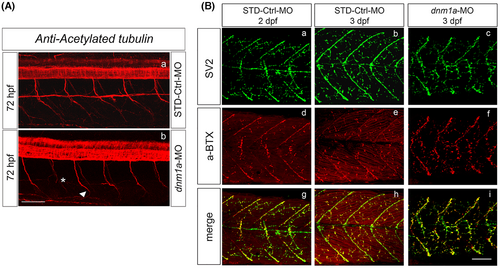
Fig. 4 Panel A. Neurite formation in dnm1a-MO-injected embryos. (A) The neurite formation on 3 dpf morphants pertaining to the C1 class was observed after acetylated tubulin staining. (a and b) Morphants at 3 dpf were compared to control embryos. In dnm1a-Mos-injected embryos, different defects in the pathfinding of motor neuron axons were observed. The morphants showed a straight ventral trajectory of CaP axons (78% of injected embryos, n = 42) compared to control embryos (n = 40). Fewer morphants (14.3% of injected embryos, n = 42) showed axons that stopped to elongate (asterisk), and an atypical curve of the terminal portion (arrowhead). Scale bar 100 μm. Panel B. Presynaptic vesicles (SV2) and AChRs (α-BTX) in dnm1a-MO-injected embryos. (a–i) In order to visualize NMJs under a confocal microscope, morphants and control embryos were double-stained with SV2 and α-bungarotoxin. (i) The merged signal observed in morphants suggested that the axonal projections were associated correctly with the cluster of AChR. (d–f) The number of NMJs did not appear to be different in the two experimental groups. The distribution pattern of AChR and NMJs appeared slightly altered in morphants compared with controls. This was explained as a secondary effect of the strong muscular defects. Scale bar 100 μm.
Image
Figure Caption
Figure Data
Acknowledgments
This image is the copyrighted work of the attributed author or publisher, and
ZFIN has permission only to display this image to its users.
Additional permissions should be obtained from the applicable author or publisher of the image.
Full text @ J. Neurosci. Res.

