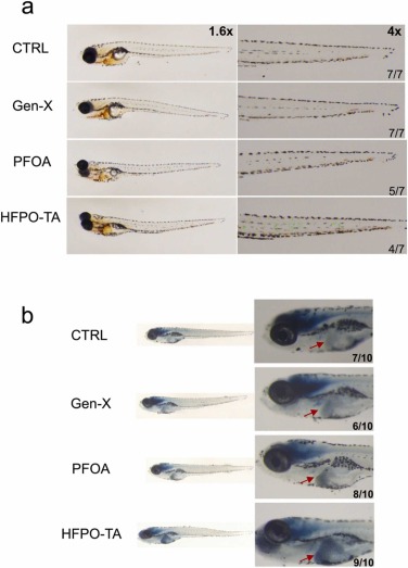
Fig. 1 Representative images of O-Dianisidine staining (a) and Sudan black B staining (b) in zebrafish after 72 hpe exposure to PFOA and alternatives. O-Dianisidine stains mature erythrocyte to reflect abnormalities in the circulatory system, and the green arrows and circles indicates a positive stain, which is dark red. Among them, PFOA, Gen-X was relatively normal, while the HFPO-TA group showed significant erythrocyte retention. n = 7, the lower right corner indicates the number of cases with the same representative image. Sudan black B staining was mainly observed for neutrophil aggregation. The figure shows the aggregation of neutrophils in all three exposure groups, and the red arrows indicate the aggregation in the liver region. n = 10, the lower right corner indicates the number of cases with the same representative image.
Image
Figure Caption
Figure Data
Acknowledgments
This image is the copyrighted work of the attributed author or publisher, and
ZFIN has permission only to display this image to its users.
Additional permissions should be obtained from the applicable author or publisher of the image.
Full text @ Ecotoxicol. Environ. Saf.

