IMAGE

Fig. 3.

ID
ZDB-IMAGE-231018-33
Source
Figures for Ali et al., 2023
Image
Figure Caption

Fig. 3.

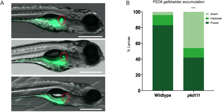
pkd1l1 knockout alters hepatobiliary function. (A) Representative images of PED6 uptake in larvae showing typical normal (top), faint (middle) and absent (bottom) fluorescence (red circles) in gallbladders. Scale bars: 500 µm. (B) Quantification of PED6 uptake showed an increase number of absent gallbladders in pkd1l1 mutant (n=145) compared to wild-type (n=84) larvae (***P<0.001; χ2 test), reflecting defects in biliary function.

Figure Data
Acknowledgments
This image is the copyrighted work of the attributed author or publisher, and ZFIN has permission only to display this image to its users. Additional permissions should be obtained from the applicable author or publisher of the image. Full text @ Dis. Model. Mech.