IMAGE

Figure 2

ID
ZDB-IMAGE-231016-4
Genes
Antibodies
Source
Figures for Hodges et al., 2023
Image
Figure Caption

Figure 2

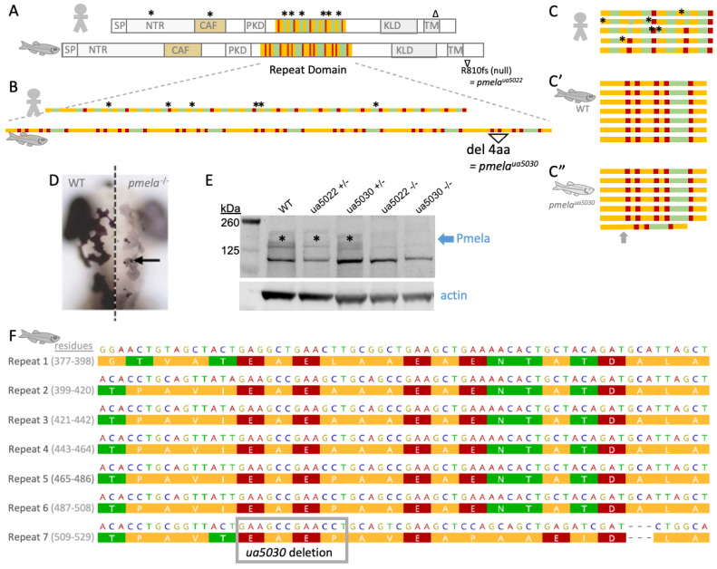
A zebrafish model of pigmentary glaucoma engineered via subtle mutation to the repeat region of the PMEL homolog that forms functional amyloid in pigment cells. (A) Homologs of PMEL protein in human and zebrafish are strikingly similar with a full complement of shared domains, including a repeat domain that contributes to making functional amyloid. Zebrafish Pmela protein is ~25% longer than human PMEL. The repeat domain in PMEL is homologous between human and zebrafish, though the details of which amino acids comprise each repeat are different; colours in panels (AC) present a simplified schematic of amino acid properties (polarity) in these repeats, with detailed views in panel F and Figure S1. Symbols * and ∆ above human PMEL indicate positions of pigmentary glaucoma patient variants (details in Figure 1), including an enrichment of missense mutations in the repeat domain [1]. (A) Previously engineered zebrafish mutant pmelaua5022 creates a premature stop codon in the N-terminus of zebrafish Pmela, and this is a null allele likely due in part to nonsense-mediated decay [1]. (B,C) Human vs. zebrafish repeat domains (simplified to emphasize repetitive aspects; see Figure S1 for details of exact amino acid content). Here, we engineered a zebrafish mutant ua5030 with an in-frame 12 bp deletion leading to a predicted loss of only 4 residues (“del 4aa”). Panel (C) presents the same protein sequences as panel B but reconfigured to emphasize the repetitive nature of the PMEL repeat domain. Human PMEL contains 5 repeats of 26 amino acids (C), in comparison to zebrafish Pmela (C’), which has 7 repeats of 22 amino acids. Akin to the non-synonymous mutations in pigmentary glaucoma patients (*), the deletion in Pmelaua5030 (C”) is predicted to subtly disrupt the overall protein but alter residues that show a rigidly repetitive character. (D) Pigment defect in larval pmela mutant zebrafish. Dorsal view, anterior at top, merge of wildtype (WT) and mutant larvae to allow side-by-side comparison at dorsal midline (dotted line). (E) Homozygous zebrafish pmela mutant larvae lacked detectable Pmela protein (both alleles). Pmela abundance was perhaps reduced in heterozygous pmela+/ua5022 but appeared similar to WT in heterozygous pmela+/ua5030 larvae. Custom Anti-Pmela antibody was raised against the repeat domain of zebrafish Pmela. (F) Zebrafish Pmela repeat region (NP_001038795.1 residues 377-529) with amino acid properties colour-coded by polarity and stacked to emphasize the rigidity of repeat identity (repeats R1 to R7 are listed alongside their residue numbering). Mutation pmelaua5030 deletes 12 bp to create a 4-residue in-frame deletion. SP = signal peptide; NTR = N-terminal region; CAF = core amyloid fragment; PKD = polycystic kidney domain; KLD = Kringle-like domain; TM = transmembrane domain.

Figure Data
Acknowledgments
This image is the copyrighted work of the attributed author or publisher, and ZFIN has permission only to display this image to its users. Additional permissions should be obtained from the applicable author or publisher of the image. Full text @ Int. J. Mol. Sci.