Image
Figure Caption

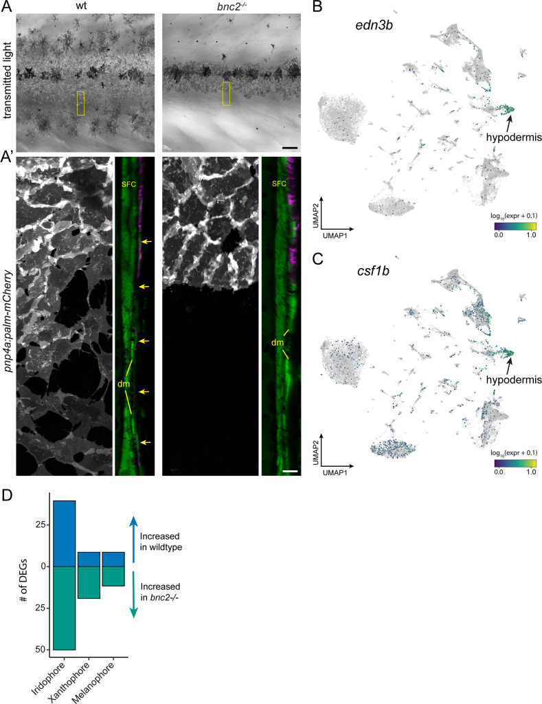
Figure 8—figure supplement 1. Hypodermis supports pigment cells.

(A) Iridophores and melanophores in wild-type and bnc2 mutant skins highlighting the relative dearth of pigment cells in the mutant. (A’) Super-resolution images of boxed region in (A), iridophores expressing pnp4a:palm-mCherry and orthogonal projection showing both iridophores and dermal cells expressing ET37:EGFP. bnc2 mutant dermis is bounded by dermal mesenchyme (dm) and lacks the thin, confluent hypodermis seen in wild-type individuals (arrows). (B, C) UMAP visualizations of pigment cell trophic factors, edn3b and csf1b. Compare to cell types illustrated in Figure 1C. (D) bnc2-dependent loss of hypodermis is associated with a greater number of differentially expressed genes (DEGs) in iridophores than in other pigment cell types. Scale bars, 100 μm (A), 10 μm (A’).

Acknowledgments
This image is the copyrighted work of the attributed author or publisher, and ZFIN has permission only to display this image to its users. Additional permissions should be obtained from the applicable author or publisher of the image. Full text @ Elife