Image
Figure Caption
Fig. 1
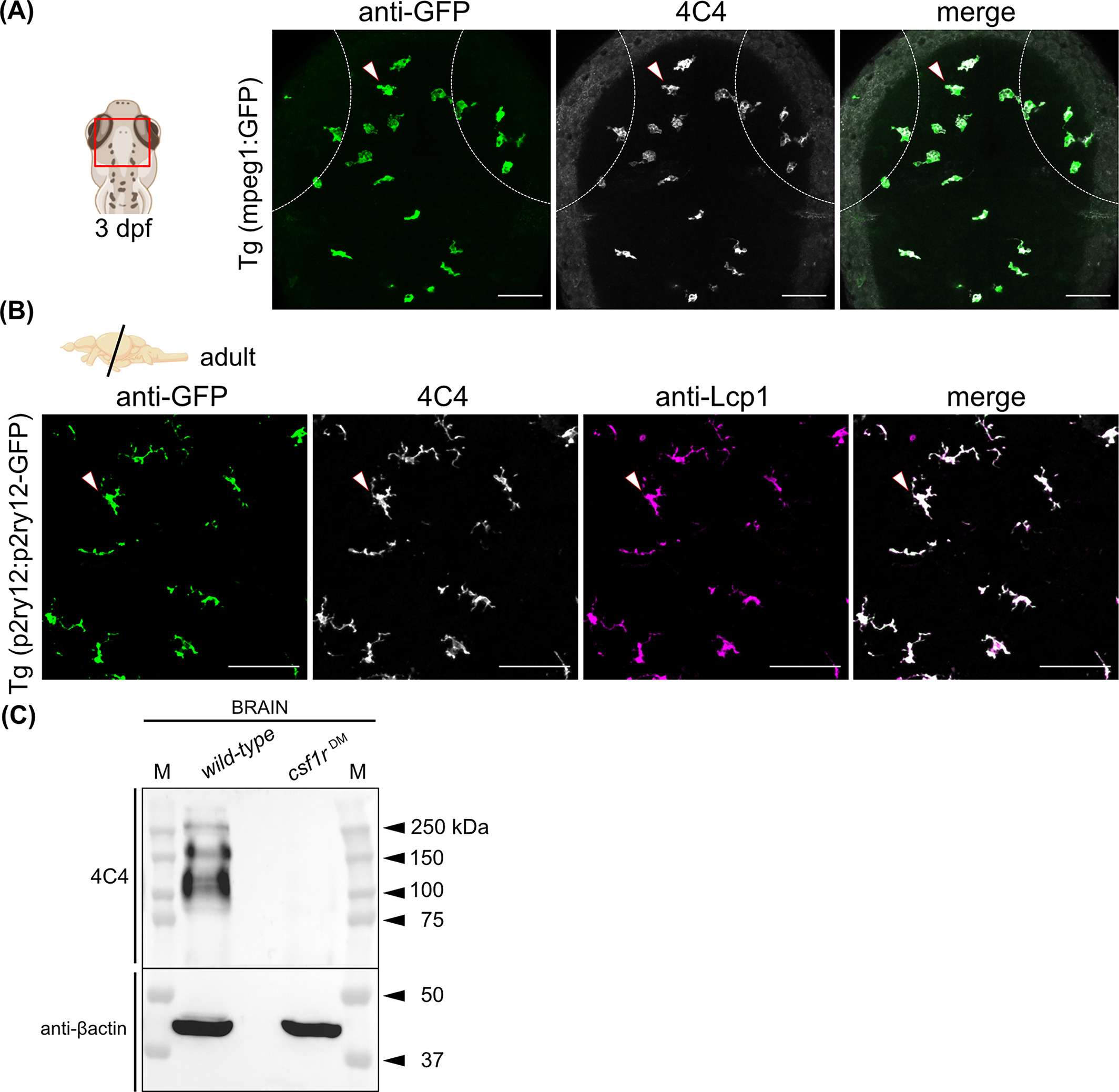
The 4C4 antibody labels microglial cells in the embryonic and adult zebrafish brain. (A) Dorsal view (red rectangle) of the optic tectum of a Tg(mpeg1:GFP) embryo at 3 dpf coimmunostained with the 4C4 antibody. Anti-GFP (green), 4C4 (gray), and merge of the two channels are shown. Dashed lines represent the eye edges. Images were taken using a 25× water-immersion objective. (B) Immunofluorescence on transversal brain sections (14 μm) from adult Tg(p2ry12-p2ry12:GFP) zebrafish co-immunostained with 4C4 and anti-Lcp1 (L-plastin) antibodies. Anti-GFP (green), 4C4 (gray), anti-Lcp1 (magenta), and merge of the three channels. Images were taken using a 20× objective. Images in (A) and (B) correspond to orthogonal projections and the white arrowheads point to microglial cells. Scale bars 50 μm. dpf, days postfertilization. (C) Detection of the 4C4 target protein by western blot. Protein lysate from a wild-type and a csf1rDM mutant adult brain. Βactin was used as a loading control. M, protein marker
Figure Data
Acknowledgments
This image is the copyrighted work of the attributed author or publisher, and
ZFIN has permission only to display this image to its users.
Additional permissions should be obtained from the applicable author or publisher of the image.
Full text @ Dev. Dyn.

