
|
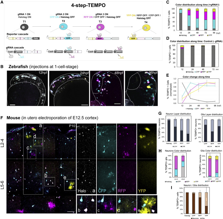
Fig. 6 Extending the color cascade to increase temporal resolution with 4-step-TEMPO
(A) Two modified TEMPO transgenes enable the ordered activation of a 4-reporter sequence (Halotag-CFP-RFP-YFP, T, temporal windows) by a parallel conditional gRNA cascade.
(B) Co-injection of two integrative ubiquitous constructs (4-step-TEMPO and Cas9-gRNA switches) into zebrafish embryos and imaging between 12 and 96 hpf. Scale bars, 25 μm.
(C and D) Changes in TEMPO color distribution (percentage of fluorescent cells for each color reporter along time).12 h, n = 4; 24 h, n = 10; 48 h, n = 8; 72 h, n = 6; 96 h, n = 6, error bars represent SEM. In the absence of the trigger gRNA (D), we observe a proportion of RFP+ and YFP+ cells (<2.5%), probably due to the leakiness of the gRNA switches (gRNA2 and gRNA3) in the absence gRNA1.
(E) Normalization of the data shown in (C) to the maximum fluorescence for each reporter along time.
(F) P10 mouse brain sections electroporated with the two modified 4-step-TEMPO constructs at E12.5. Dashed line: limit between upper (L2-4) and lower layers (L5-6). Insets: TEMPO+ lower layer neurons expressing Halotag and CFP (B) or RFP+ astrocytes (C), CFP+ and RFP+ upper layer neurons (D), or YFP+ astrocytes (E).
(G) Layer distribution of TEMPO+ neurons (left) or glia (right).
(H) Percentage of neurons (left) or glia (right) expressing each TEMPO reporter in the upper (L2-4) or lower (L5-6) layers.
(I) Neuron and glia ratio for each reporter. Data are represented as percent ± SEM. n = 3, Scale bars, 100 μm.