IMAGE

Fig. 1.

ID
ZDB-IMAGE-230609-37
Genes
Source
Figures for Lelièvre et al., 2023
Image
Figure Caption

Fig. 1.

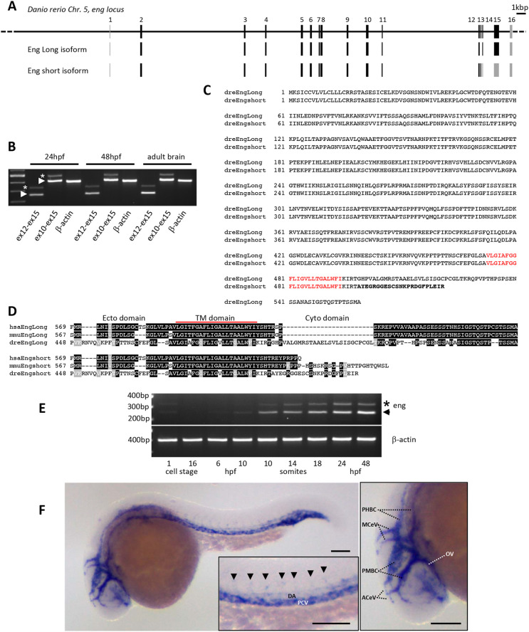
Characterization of zebrafish endoglin chromosomal organization, transcripts and expression pattern. (A) Zebrafish endoglin gene exon-intron structure on chromosome 5. Coding exons (black) and non-coding exons (gray) are shown. (B) RT-PCR analysis of endoglin variants. Endoglin long (arrowheads) and short (asterisks) isoform messenger expression in embryo and adult tissue (brain). (C) Protein alignment of zebrafish Endoglin isoforms. Alternative amino acids (aa) of Endoglin short isoform are in bold. Transmembrane aa are in red. (D) Alignment of human, mouse and zebrafish Endoglin short isoform C-terminal region. Identical (black) and similar (gray) aa are shown. TM, transmembrane. (E) RT-PCR analysis of endoglin variants during zebrafish development. Endoglin short isoform (asterisk) and Endoglin long isoform (arrowhead) are shown. hpf, h postfertilization. (F) Left: whole-mount in situ hybridization using endoglin antisense riboprobe on 24 hpf wild-type embryo. Inset: close-up of the trunk region showing differential endoglin expression between the dorsal aorta (DA) and the posterior cardinal vein (PCV). Arrowheads indicate intersegmental vessels. Right: close-up of the head region showing mostly venous endoglin expression. ACeV, anterior cerebral vein; MCeV, middle cerebral vein; PHBC, primordial hindbrain channel; PMBC, primordial midbrain channel; OV, optic vein. Scale bars: 100 µm.

Figure Data
Acknowledgments
This image is the copyrighted work of the attributed author or publisher, and ZFIN has permission only to display this image to its users. Additional permissions should be obtained from the applicable author or publisher of the image. Full text @ Dis. Model. Mech.