
Image
|
Figure Caption
Figure 1
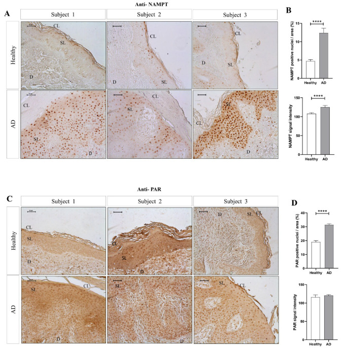
The amount of NAMPT protein and PARylation increased in lesional skin of AD patients. Representative images and analysis of biopsy sections from healthy (n = 10) and AD (n = 6) skin biopsies immunostained with anti-NAMPT (A,B) and anti-PAR (C,D). The mean ± SEM of each group is shown. p values were calculated using the nonparametric Mann–Whitney test, **** p ≤ 0.0001. Scale bar is 50 μm in all panels. CL: cornified layer; D: dermis; SL: spinous layer.
Acknowledgments
This image is the copyrighted work of the attributed author or publisher, and
ZFIN has permission only to display this image to its users.
Additional permissions should be obtained from the applicable author or publisher of the image.
Full text @ Int. J. Mol. Sci.