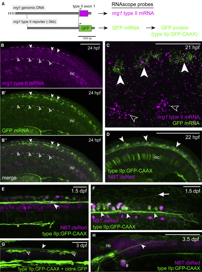
Fig. 4 Unmyelinated RB sensory neurons express nrg1 type II
(A) Diagram indicating location of 3 kb regulatory element and reporter construct design. Colored rectangles indicate coding regions; open rectangles indicate UTRs. Arrows indicate transcriptional start sites.
(B) Endogenous nrg1 type II mRNA detected by RNAScope in situ hybridization at 24 hpf. White arrowheads mark expression in dorsal spinal cord. Black arrowheads mark expression in the region of motor neurons in the ventral spinal cord. Expression is also evident in the notochord (nc). Dotted lines mark the dorsal and ventral extent of the spinal cord in the posterior trunk and anterior tail. Scale bar, 100 μm. (B’) In the same animal as (B), GFP mRNA from the nrg1 type II reporter transgene was also detected by RNAScope in situ hybridization. (B’’) Merge of (B) and (B’). Co-expression of GFP mRNA with endogenous nrg1 type II mRNA is observed in both the dorsal spinal cord and notochord. Representative images from five replicate experiments.
(C) Co-expression of endogenous nrg1 type II mRNA and GFP mRNA from the nrg1 type II reporter transgene. White arrowheads indicate putative cell bodies of three GFP-expressing Rohon-Beard (RB) sensory neurons, which also express endogenous nrg1 type II mRNA. Black arrowheads indicate two areas of endogenous nrg1 type II mRNA expression in the region of motor neurons. Scale bar, 50 μm.
(D) GFP from the nrg1 type II reporter is expressed prominently in early-born Rohon-Beard (RB) neurons in the dorsal spinal cord (arrowheads; colocalized with the pan-neuronal marker NBT:dsRed45)and transiently in the notochord (nc) at 22 h postfertilization. Scale bar, 200 μm.
(E) Large, GFP-expressing RB neurons send axons (arrowhead) anteriorly and posteriorly via the dorsal longitudinal fasciculus (DLF). Reporter expression also occurs in the posterior lateral line nerve (asterisk), which grows posteriorly at 1.5 dpf. Scale bar, 50 μm.
(F) The nrg1 type II reporter is expressed in neurons with large cell bodies and sensory arbors (arrow) characteristic of RB neurons at 1.5 days post fertilization (1.5 dpf). White arrowheads indicate the DLF as in (E). Scale bar, 50 μm.
(G) GFP-expressing axons from RB neurons (white arrowhead) are near oligodendrocytes (black arrowheads) in the dorsal spinal cord. Scale bar, 50 μm.
(H) GFP-expressing axons in the DLF (arrowhead) project anteriorly into the hindbrain (hb). GFP-expressing lateral line nerve descends from the posterior lateral line ganglion (#). Scale bar, 200 μm.

