Figure 1
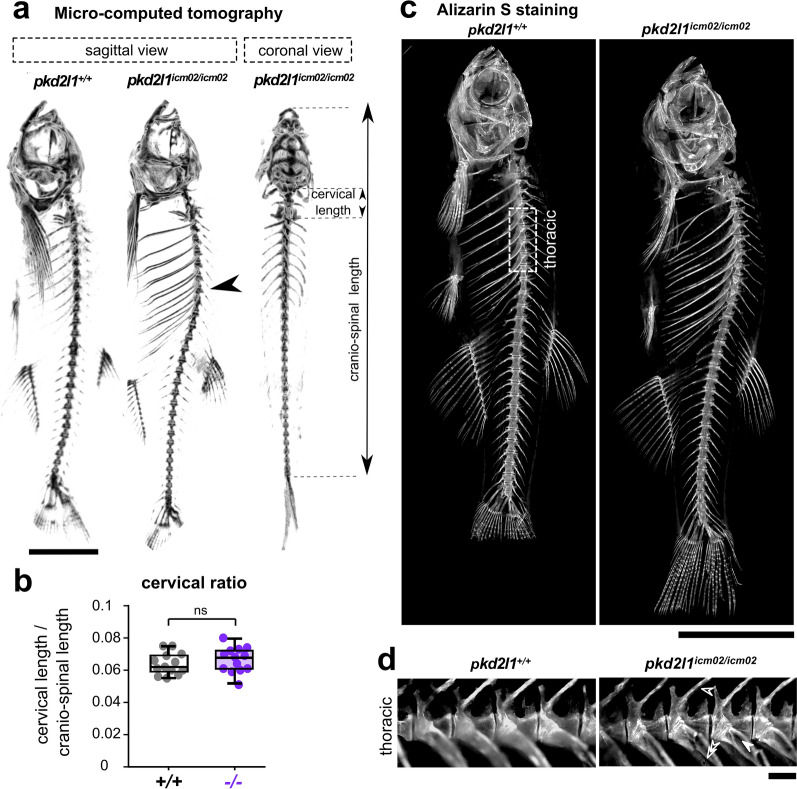
pkd2l1 null mutants develop during adulthood an idiopathic hyper-kyphosis of the thoracic spine. (a) 3D micro-computed tomography reconstruction images of a 18 months-old wild-type (pkd2l1+/+, left sagittal view) and a pkd2l1icm02/icm02 mutant sibling (middle, sagittal view; right, frontal view). pkd2l1icm2/icm02 mutants exhibit a thoracic hyper-kyphosis of the spine (arrowhead) but show no deformity in the coronal plane. Scale bar: 0.5 cm. (b) Distribution at 18 months-old of the cervical ratio, defined as the cervical length divided by the cranio-spinal length, in wild-type (grey, + / + , n = 13) and pkd2l1icm02/icm02 mutants (purple, −/−, n = 13). Each point represents a single animal. Boxplots represent median values ± IQR. ns: p > 0.05, Mann–Whitney test. (c) Sagittal views of Alizarin S stained-skeletal preparations of 18 months-old wild-type (pkd2l1+/+, left) and a pkd2l1icm02/icm02 mutant sibling (right). Scale bar: 0.5 cm. (d) High-magnification fluorescence images of Alizarin S in the thoracic region of the spine (as defined in (c), dotted line rectangle) in a wild-type (left) and a pkd2l1icm02/icm02 sibling (right). Neural arches (empty arrowhead), hemal arches (double empty arrowhead) and ribs (solid arrowhead) are highlighted for the pkd2l1icm02/icm02 mutant. Note that in both genotypes, no vertebral dislocation, malformation or fusion are observed. Scale bar: 500 µm.

