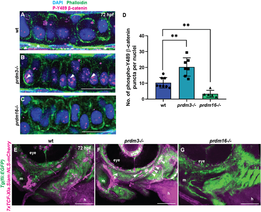
Fig. 4 Prdm3 and Prdm16 control β-catenin stabilization and localization in craniofacial chondrocytes. (A-D) Wild-type (wt), prdm3−/− and prdm16−/− zebrafish embryos were collected at 75 hpf and immunostained for nuclear β-catenin (phosphorylated tyrosine 489) and counterstained with phalloidin and DAPI (A-C). Shown are high-magnification lateral images of the palatoquadrate. Increased nuclear β-catenin (magenta) was observed in prdm3−/− (white arrowheads in B), which was significantly reduced in prdm16−/− (C). (D) Quantification of the number of β-catenin puncta across ten nuclei per individual and averaged across at least five embryos per genotype; mean±s.d. Scale bars: 50 µm. (E-G) prdm3−/− and prdm16−/− mutant lines were crossed into the Wnt reporter line Tg(7xTCF-Xla.Siam:NLS-mCherry) to assess Wnt-responsive cells. Shown are representative lateral-ventral views of 75 hpf wild-type (E), prdm3−/− (F) and prdm16−/− (G) embryos. Increased Wnt-responsive cells were identified in the pharyngeal arch tissues of prdm3−/− (F) (white arrowheads), with a dramatic decrease in Wnt-responsive cells in prdm16−/− (G) mutants compared with wild type (E). h, heart; m, mouth. Scale bars: 100 µm. **P≤0.005 (unpaired, two-tailed Student's t-test).
Image
Figure Caption
Figure Data
Acknowledgments
This image is the copyrighted work of the attributed author or publisher, and
ZFIN has permission only to display this image to its users.
Additional permissions should be obtained from the applicable author or publisher of the image.
Full text @ Development

