
|
Fig. 6
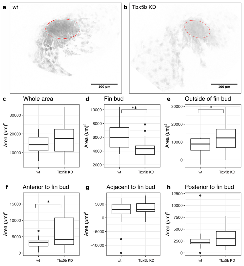
(a-b) Maximal intensity projections of representative fin buds at 30 hpf of Tg(Tbx5a::GFP) embryos. The fin bud is outlined in red. Anterior is to the left, the dorsal midline is up. Representative images are shown for both (a) uninjected and (b) Tbx5b knock-down embryos. (c-h) Measurements of the area of GFP+ expressing cells. For both conditions, n=8 embryos/16 limb buds. (c) There is no significant difference in the total area of GFP+ expressing cells between wt and Tbx5b knock-down embryos. (d) There is a significant (p<0.01) difference in the area of GFP+ expressing cells in the fin bud between wt and Tbx5b knock-down embryos. (e) There is a significant difference (p<0.05) in the area containing GFP+ expressing cells outside of the fin bud between wt and Tbx5b knock-down embryos. (f) There is a significant difference (p<0.05) in the area of GFP+ cells located rostrally to the fin bud between wt and Tbx5b knock-down embryos. There is no significant difference between wt and Tbx5b knock-downs in the area of GFP+ expressing cells located either (g) adjacent to or (h) posterior to the fin bud.
Reprinted from Developmental Biology, 481, Boyle-Anderson, E.A.T, Mao, Q., Ho, R.K., Tbx5a and Tbx5b paralogues act in combination to control separate vectors of migration in the fin field of zebrafish, 201-214, Copyright (2021) with permission from Elsevier. Full text @ Dev. Biol.