
|
Fig. 3
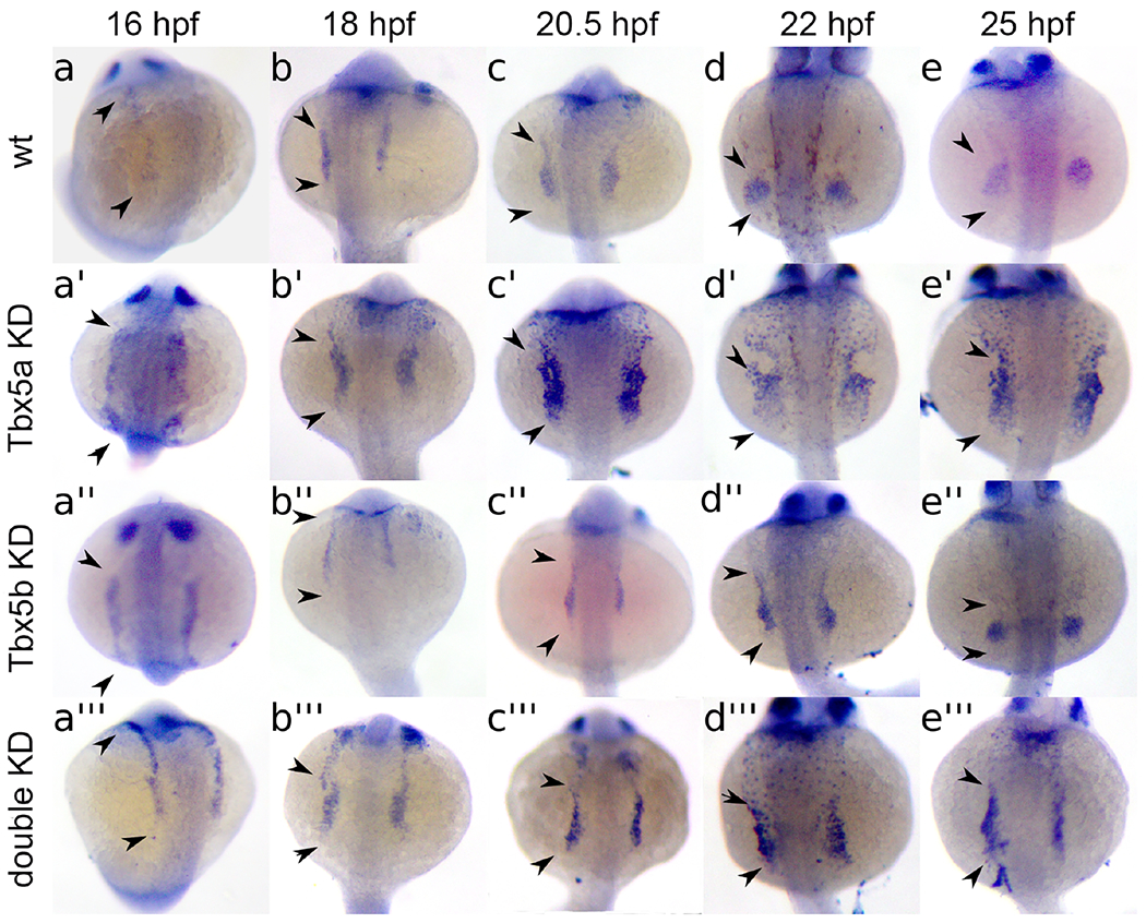
Embryos are shown in a dorsal view with anterior to the top. Embryos are at 16 hpf (a, a’, a’’, a’’’), 18 hpf (b, b’, b’’, b’’’), 20.5 hpf (c, c’, c’’, c’’’), 22 hpf (d, d’, d’’, d’’’), and 25 hpf (e, e’, e’’, e’’’). Control embryos are either uninjected wt (a) or wt injected with a control morpholino (b-e), and show a condensing in the shape of the pectoral fin field tbx5a expression to a circular shape over time. (a’-e’) Tbx5a knock-down embryos display a lack of condensing of pectoral tbx5a expression over time, leading to wider, more scattered expression pattern. (a’’-e’’) Tbx5b knock-downs display a delay in the condensing of pectoral fin bud tbx5a expression over time to a disc pattern. (a’’’-e’’’) Double knock-downs display a relatively constant expression pattern of tbx5a over time. Arrowheads in a-a’’’ indicate the extent of continuous tbx5a expression before the separation of the fin field region from more anterior LPM. Arrowheads in b-e’’’ indicate the extent of tbx5a expression in the fin field. Width of yolk approximately equals 500μm.
Reprinted from Developmental Biology, 481, Boyle-Anderson, E.A.T, Mao, Q., Ho, R.K., Tbx5a and Tbx5b paralogues act in combination to control separate vectors of migration in the fin field of zebrafish, 201-214, Copyright (2021) with permission from Elsevier. Full text @ Dev. Biol.