
|
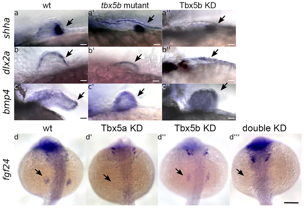
Fig. 2
(a-c’’) Lateral views with anterior to the left. Scale bars=10 μm. (a-a’’) shha (arrows) is expressed in the posterior mesenchyme of the fin bud at 32 hours post fertilization (hpf) in wt embryos (a), tbx5b mutant (a’), and Tbx5b knock-down (a’’) embryos. (b-b’’) dlx2a (arrows) is expressed in the AER of the fin bud at 32 hpf in the wt (b), tbx5b mutant (b’) and Tbx5b knock-down (b’’) embryos, (c-c’’) bmp4 (arrows) is expressed in the wt fin at 48 hpf (c). tbx5b mutant (c’) and Tbx5b knock-down (c’’) embryos, (d-d’’’) Arrows indicate the normal location of fgf24 expression in the fin field at 21 hpf. Embryos are shown in a dorsal view with anterior to the top. Scale bar =100μm. (d) fgf24 is expressed in the fin field of wildtype embryos. (d’) fgf24 is absent from the fin field of Tbx5a knock-down embryos. (d’’) fgf24 expression is decreased in the fin field of Tbx5b knock-down embryos. (d’’’) fgf24 expression is absent in double knock-down embryos.
Reprinted from Developmental Biology, 481, Boyle-Anderson, E.A.T, Mao, Q., Ho, R.K., Tbx5a and Tbx5b paralogues act in combination to control separate vectors of migration in the fin field of zebrafish, 201-214, Copyright (2021) with permission from Elsevier. Full text @ Dev. Biol.