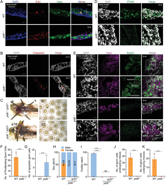
Figure 5
Blocking of germ cell self‐renewal and differentiation in pld6‐ depleted gonad. A) EdU staining of proliferating cells in wildtype and pld6 −/‐ juvenile gonads. The germ cells were marked by anti‐vasa immunostaining. N represents analyzed individual number. Scale bar: 100 µm. B) Apoptosis detection in wildtype and pld6 −/− juvenile gonads by anti‐Caspase3 immunostaining. N represents analyzed individual number. Scale bar: 100 µm. C)Morphological observation of gonads and offspring of pld6 −/− and pld6 −/−; tp53−/− double mutant. D) Detection of mitosis in wildtype and pld6 −/‐ juvenile gonads by anti‐Pcna immunostaining. N represents analyzed individual number. Scale bar: 100 µm. E) Detection of meiosis in wildtype and pld6 −/− juvenile gonads by anti‐Sycp3 immunostaining. N represents analyzed individual number. Scale bar: 100 µm. F) Statistical analysis of total number of proliferating germ cells. G) Statistical analysis of the total number of apoptotic germ cells. H) Sex ratio of wildtype, pld6−/− , and pld6 −/−; tp53−/− double mutant. I) Fertilization rates of wildtype, pld6−/− and pld6 −/−; tp53−/− double mutant. J) Statistical analysis of the total number of germ cells undergoing mitosis. K) Statistical analysis of the total number of germ cells undergoing meiosis. The data were expressed as mean ± SD. The P values in this figure were calculated by two‐sided t‐test. ***P < 0.001; ns, no significant difference.

