
|
Figure 2
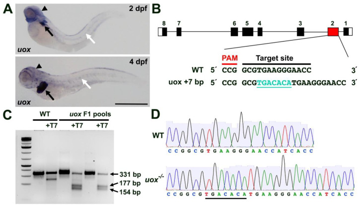
Generating a zebrafish uox mutant. (A) Expression of uox in 2 and 4 day post fertilization (dpf) larvae, as detected by WMISH (black arrows and arrowheads mark liver and head expression, respectively. White arrows mark horizontal myoseptum expression). (B) Schematic illustrating the intron (black line) and exon (black rectangles) structure of uox (with flanking UTRs as white rectangles). The position (red rectangle) of the CRISPR/Cas9 target site and the sequence are annotated, with the +7 indel mutation shown in green. (C) T7 endonuclease I assay (with and without T7 endonuclease) on pools of WT and F1 embryos. Successful editing is indicated by the cleavage products of 177 bp and 154 bp. (D) Sequencing chromatogram of amplicons from WT uox compared to a homozygous carrier of the +7 bp uox mutation (underlined). Scale bar: 500 µm.