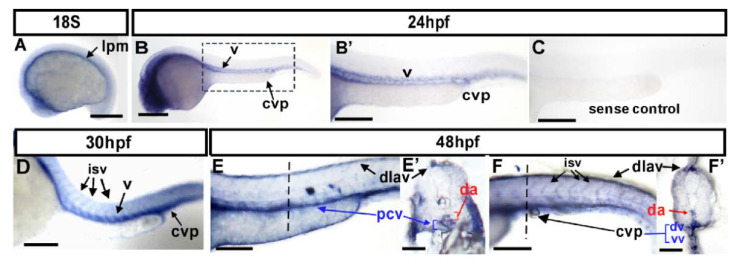
Figure 1
Expression pattern of gtpbp1l mRNA during zebrafish vessel development. (A–F) Spatiotemporal expression of gtpbp1l in the vessels (v), caudal vein plexus (CVP), intersegmental vessels (isv), and dorsal longitudinal anastomotic vessel (dlav) during development as shown at 18S, 24 hpf, 30 hpf, and 48 hpf. A cross-section taken at 48 hpf (E’,F’) clearly shows the expression of gtpbp1l in the dorsal aorta (da), posterior vein (pcv), and CVP. (A) At the 18 somite (18S) stage, gtpbp1l expression is in the lateral plate mesoderm (lpm). (B,B’) At 24 hpf, gtpbp1l is expressed in the vessels (v) and caudal vein plexus (CVP). (B’) is an enlargement of (B). (C) The gtpbp1l sense probe served as a negative control. (D) At 30 hpf, gtpbp1l expression can be observed in the vessels, isv, and CVP (E,E’,F,F’) At 48 hpf, gtpbp1l is expressed continuously in the vessels, isv, dlav, and CVP at the region of the trunk (E) and tail (F). (E’,F’) Cross-sections of embryos from (E,F) show that gtpbp1l is expressed in the dorsal aorta (da), posterior cardinal vein (pcv), dorsal vein (dv), and ventral vein (vv) of the CVP. Scale bars are 200 µm.

