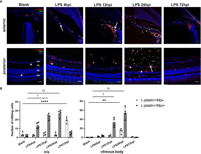
Distribution of immune cells during EIU inflammation process in zebrafish. (A) L-plastin (magenta), Mpx (green), and DAPI (blue) staining was seen in cryosections at 0 hpi (blank control), 4 hpi, 12 hpi, 24 hpi and 72 hpi of LPS. ONL, outer nuclear layer, INL, inner nuclear layer, IPL, inner plexiform layer, GCL, ganglion cell layer. Asterisk: L-plastin+/Mpx- cell. White arrow: L-plastin+/Mpx+ cell. Scale bar: 20μm. (B) Quantification of immune cells in the iris and vitreous body at each time point (mean ± SD; *P < 0.05, **P < 0.01, ****P < 0.0001, the number of L-plastin+/Mpx- cells were compared; †P < 0.05, ††P < 0.01, number of L-plastin+/Mpx+ cells were compared, one-way ANOVA).
Acknowledgments
This image is the copyrighted work of the attributed author or publisher, and
ZFIN has permission only to display this image to its users.
Additional permissions should be obtained from the applicable author or publisher of the image.
Full text @ Front Immunol
Your Input Welcome
Thank you for submitting comments. Your input has been emailed to ZFIN curators who may contact you if
additional information is required.
Oops. Something went wrong. Please try again later.