IMAGE

Figure 2

ID
ZDB-IMAGE-221218-15
Source
Figures for Martin et al., 2022
Image
Figure Caption

Figure 2

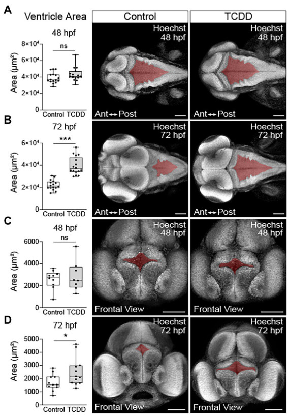
TCDD exposure increases brain ventricle size. To determine the impact of TCDD exposure on ventricular development, the hindbrain (A,B) and forebrain (C,D) ventricles were measured in embryos ranging in age from 48 to 52 hpf (A&C) and larvae from 72 to 76 hpf (B,D). TCDD exposure significantly increased hindbrain and forebrain ventricle size at 72 hpf (B,D) with non-significant trends observed early in development. Dorsal 10x magnification (A,B) and frontal (C,D) 20x confocal micrographs of zebrafish embryos and larvae stained with Hoechst. N = 15–17 fish/group hindbrain across 3 replicates; n = 8–12 fish/group forebrain across replicates. Anterior (Ant) is left, posterior (Post) is right in confocal micrographs (A,B). Scale bar = 100 μm. Statistical significance determined by Welch’s t-test, *p < 0.05, ****p < 0.0001.

Acknowledgments
This image is the copyrighted work of the attributed author or publisher, and ZFIN has permission only to display this image to its users. Additional permissions should be obtained from the applicable author or publisher of the image. Full text @ Front. Mol. Neurosci.