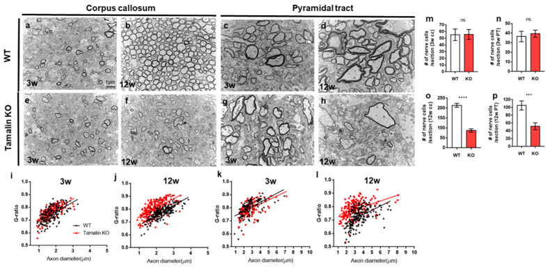
Figure 5 Tamalin KO induces nerve degeneration and hypomyelination in the postembryonic mouse brain. (a–h) TEM images displaying transverse sections of the corpus callosum (a,b,e,f) and pyramidal tract (c,d,g,h) at 3 weeks (a,e,c,g) and 12 weeks (b,f,d,h) in wildtype and tamalin KO mice. (i–l) The g-ratio of myelinated axons in the corpus callosum (i,j) and pyramidal tract (k,l) of wildtype and tamalin KO zebrafish. Unpaired t test was used to compare means from each animal. Each g-ratio was from 100 myelinated axons in eight sections of four mice each (i: p = 0.0535, k: p = 0.0579, j,l: **** p < 0.0001). (m–p) Quantification of the number of nerve cells in the corpus callosum (m,o) and pyramidal tract (n,p) of wildtype and tamalin KO mice (**** p < 0.0001, *** p < 0.001). ns; no significance. Scale bars: (a–h): 1 μm. KO, knockout; TEM, transmission electron microscopy.
Image
Figure Caption
Acknowledgments
This image is the copyrighted work of the attributed author or publisher, and
ZFIN has permission only to display this image to its users.
Additional permissions should be obtained from the applicable author or publisher of the image.
Full text @ Int. J. Mol. Sci.

