
|
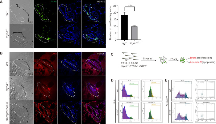
Fig 4
(A) Cell proliferation in the intestines was detected by PCNA immunofluorescence staining (green signal) of the tissue sections from the WT and mycn mutant embryos. The statistical analysis of the proliferating cells of the sections of intestine in the right. (B) Apoptosis in the intestines was detected via TUNEL assay (red signal) for the tissue sections from the WT, mycn mutant, and camptothecin-treated (as positive control) embryos. Sections were cut along the transverse plane. Dotted lines indicate the intestine positions. (C) Schematic representation of the experimental workflow of the flow cytometry analysis. (D) Cell proliferation in the intestines was detected by BrdU incorporation assay. (E) Apoptosis in the intestines was detected by APC-Annexin V staining. Embryos used in the flow cytometry analysis were descendants of ET33J1: EGFP reporter line crossed with WT or mycn mutants (D and E). Scale bar: 50 μm. PCNA, proliferating cell nuclear antigen; WT, wild-type.