
|
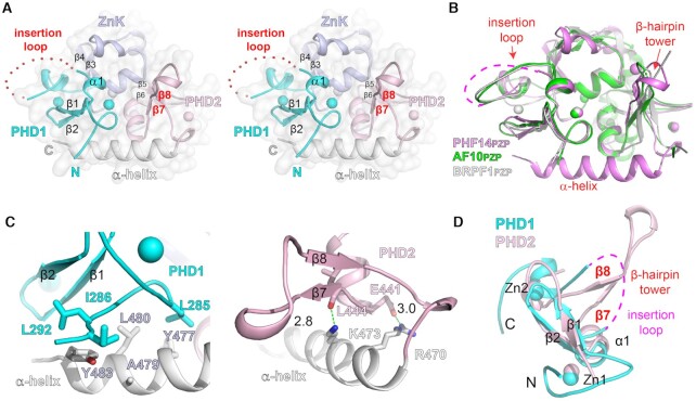
Fig. 2
Crystal structure of PHF14PZP in a peptide-free state. (A) Wall-eye stereo view of the overall structure of free PHF14PZP. PHD1 (cyan), ZnK (light blue), PHD2 (light pink), and α-helix (white) are shown as ribbons. Zinc ions are depicted as spheres. Red dotted lines indicate the invisible insertion loop. Key secondary structural elements are labeled. (B) Structural alignment of PHF14PZP (magenta), AF10PZP (green) and BRPF1PZP (white). (C) Interactions between the C-terminal α-helix with PHD1 (left) and PHD2 (right). Key residues are shown as sticks. Hydrogen bonds are shown as green dashes with distances labeled in Å. (D) Structural alignment of PHF14PHD1 (cyan) and PHF14PHD2 (light pink). Note that the insertion loop is topologically equivalent to the β-hairpin tower.