Fig. 5
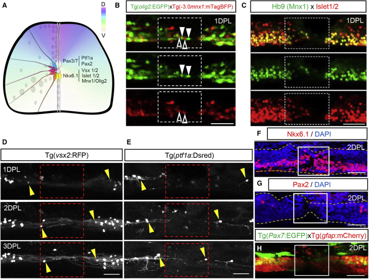
Figure 5. Molecular characteristics of neurons rapidly recruited to the injury (A) Schematic cross section of the spinal cord showing dorsoventral domains. (B) Maximum projection side views of olig2:EGFP (green) and mnx1:mTagBFP (red) fish at 1 DPL showing recruitment of ventral cell lineages to the injury. Arrowheads downward show olig2:EGFP+ cells and arrowheads upward show mnx1:mTagBFP+ neurons. (C) Hb9 (Mnx1) and Islet1/2 immunohistochemistry showing recruitment of ventral cells to the injury at 1 DPL. (D) Sagittal view showing slow recruitment of interneurons expressing vsx2:RFP to the lesion site between 1–3 DPL. (E) Sagittal view showing marginal recruitment of dorsal inhibitory sensory neurons using the ptf1a:DsRed reporter between 1–3 DPL. (F and G) Sagittal view showing abundant Nkx6.1+ but no Pax2+ immunolabelled cells at the lesion site 2 DPL. Spinal cord boundaries are outlined in yellow. (H) Maximum projection of pax7a:EGFP labeling dorsal cell lineages at 2 DPL. The lesion site is outlined in white and is devoid of GFP+ cells. Scale bar, 50 μm.
Reprinted from Developmental Cell, 56, Vandestadt, C., Vanwalleghem, G.C., Khabooshan, M.A., Douek, A.M., Castillo, H.A., Li, M., Schulze, K., Don, E., Stamatis, S.A., Ratnadiwakara, M., Änkö, M.L., Scott, E.K., Kaslin, J., RNA-induced inflammation and migration of precursor neurons initiates neuronal circuit regeneration in zebrafish, 2364-2380.e8, Copyright (2021) with permission from Elsevier. Full text @ Dev. Cell

