Fig. 3
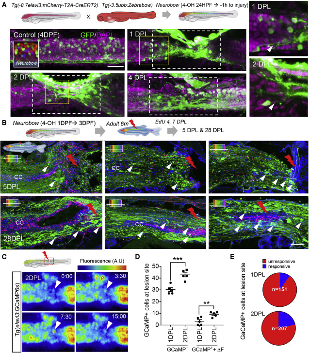
Figure 3. Tissue remodeling and cell migration drive the early phase of spinal cord regeneration (A) Recombination of neurons during embryonic development using Neurobow. Embryos were treated with 4-OH from early neurogenesis (24 hpf) until 1 h before injury. Maximum projection images at the injury site at 1, 2, and 4 DPL showing embryonically produced neurons (green) and their processes (white arrowhead) recruited rapidly to the injury site. (B) SCL in adult Neurobow fish at 6 months (embryonically recombined as in A). Fish received EdU 4 and 7 DPL and were analyzed at 5 and 28 DPL. Horizontal sections at different levels of the spinal cord demonstrating recruitment of embryonically produced neurons (green) to the lesion site (white arrowhead) in addition to the non-overlapping newly produced cells labeled with EdU (Red). (C) Snapshot at 2 DPL of a time-lapse movie showing the first ΔF active neurons at the lesion site, white arrowhead, (Video S3). Times in S:ms. (D and E) Quantification of neurons and active neurons (ΔF) at the lesion site between 1–2 DPL (n = 5 for each time point). The proportion of active neurons is low at 1 DPL (9.8%) and increases at 2 DPL (21.4%). Error = SEM, ∗∗p < 0.01, ∗∗p < 0.001.
Reprinted from Developmental Cell, 56, Vandestadt, C., Vanwalleghem, G.C., Khabooshan, M.A., Douek, A.M., Castillo, H.A., Li, M., Schulze, K., Don, E., Stamatis, S.A., Ratnadiwakara, M., Änkö, M.L., Scott, E.K., Kaslin, J., RNA-induced inflammation and migration of precursor neurons initiates neuronal circuit regeneration in zebrafish, 2364-2380.e8, Copyright (2021) with permission from Elsevier. Full text @ Dev. Cell

