Fig. 1
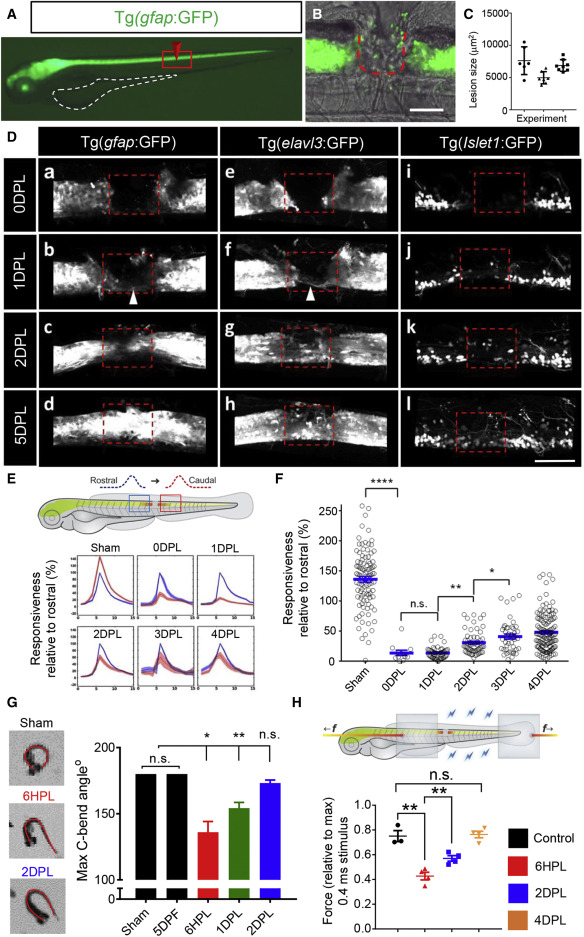
Figure 1. Rapid cellular, circuit, and functional recovery after spinal cord injury (A) Lateral view of a gfap:GFP larva at 3 dpf. Yolk sac outlined in white; the red box shows the field of view in (B). (B) Surgery at the level of the anal pore entirely ablates the spinal cord, scale bar, 100 μm. (C) Quantification of the lesion shows high reproducibility of the injury size between experiments. (D) Live imaging of glial gfap:GFP and neuronal elavl3:GFP, isl1:GFP transgenic reporters demonstrating rapid regenerative response of diverse cell types following injury. The lesion site is indicated by dashed box. At 1–2 DPL, neuronal and glial bridging and a small number of neurons within the lesion site. At 5 DPL, the neuronal and glial tissue architecture was largely restored. (E) Quantification of GCaMP6s fluorescence (ΔF) rostral (blue) and caudal (red) to the site of injury at 0–4 DPL. n was; uninjured = 9, 0DPL = 3, 1-4DPL = 5. (F) Neural activity downstream of injury represented as % relative to the equivalent upstream response over time. (G) Dorsal view of the evoked C-bend angle. Complete 180° response in intact larvae, reduced in 6HPL (24%), 1 DPL (14%) larvae and recovered by 2 DPL. (H) Schematic of muscle force recording. Force was measured between the clips (gray boxes). A significant improvement in force production by 2 DPL (0.4 ms pulse stimulus), n = 4–20 larvae, error = SEM (standard error of the mean), n.s., not significant, ∗p < 0.05, ∗∗p < 0.01, ∗∗∗∗p < 0.0001. Dorsal is up and rostral is left in all images.
Reprinted from Developmental Cell, 56, Vandestadt, C., Vanwalleghem, G.C., Khabooshan, M.A., Douek, A.M., Castillo, H.A., Li, M., Schulze, K., Don, E., Stamatis, S.A., Ratnadiwakara, M., Änkö, M.L., Scott, E.K., Kaslin, J., RNA-induced inflammation and migration of precursor neurons initiates neuronal circuit regeneration in zebrafish, 2364-2380.e8, Copyright (2021) with permission from Elsevier. Full text @ Dev. Cell

