
Image
|
Figure Caption
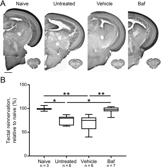
Fig. 11
Fig. 11. Quantification of tectal reinnervation via anterograde biocytin tracing six days post optic nerve damage in untreated, vehicle-treated or bafilomycin A1-treated fish. (A) Representative images and semi-quantitative analysis (B) of the tectal area covered by RGC axon terminals show that bafilomycin A1 has a positive effect on axonal regeneration as more axons re-entered the optic tectum after autophagy inhibition, compared to untreated or vehicle-treated control fish. Scale bar = 200 µm. N = 6–7, except naive (3). Baf, bafilomycin A1; dpi, days post-injury.
Acknowledgments
This image is the copyrighted work of the attributed author or publisher, and
ZFIN has permission only to display this image to its users.
Additional permissions should be obtained from the applicable author or publisher of the image.
Full text @ Neuroscience