
Image
|
Figure Caption
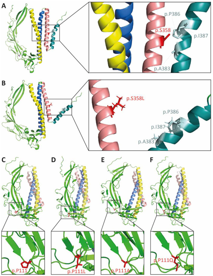
Fig. 1
The p.S358L variant changes protein conformation of TMEM43. In silico modeling of TMEM43 tertiary structure of wild-type (A,C) and proteins with the corresponding missense mutations (B) p.S358L, (D) p.P111L, (E) p.P111A, and (F) p.P111Q. Yellow indicates transmembrane domain (TMD) 1, blue indicates TMD2, rose indicates TMD3, and turquoise indicates TMD4. The residue of interest is indicated in red (p.S358 in A and B; p.P111 in C–F).
Acknowledgments
This image is the copyrighted work of the attributed author or publisher, and
ZFIN has permission only to display this image to its users.
Additional permissions should be obtained from the applicable author or publisher of the image.
Full text @ Int. J. Mol. Sci.