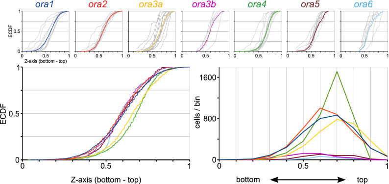
Fig. 5
ora4-expressing cells show preferential expression in the top of the olfactory organ. Quantitative assessment of distribution of ora-expressing neurons along the vertical z-axis (height-within-the-organ). Height within the olfactory organ was quantified as section number in a series of horizontal sections, and normalized to the total number of sections containing sensory epithelium, using the same set of cells, for which laminar height was determined. Relative height-within-the-organ ranges from 0 (bottommost section) to 1 (top section, near to the opening of the bowl-shaped olfactory organ). Upper row: The resulting distributions are shown unbinned (ECDF). Light grey curves represent the distribution for individual olfactory organs, colored curves represent the cumulative distribution for the respective genes. The color code for the ora genes is the same as in other figures to facilitate comparisons between different positional parameters. Lower row: Overlay of the distributions shown individually in the upper row, both as ECDF (left panel) and histogram (right panel). Due to lower resolution (all cells within one 10 µm section are assigned to a single z value) the ECDF curves appear ragged.

