Image
Figure Caption
Fig. 2
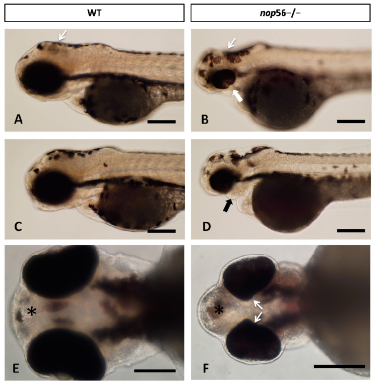
(A–D) Lateral views of WT and nop56−/− larvae (3.5 dpf), showing coloboma (white thick arrow in (B)) and jaw malformation (black arrow in (D)) in the homozygous fish. Note also differences in the midbrain and cerebellum (thin white arrows). (E,F) Ventral view of WT and nop56−/− fish showing malformation of the eye (arrows) and forebrain (asterisk). Scale bar: 125 μm.
Figure Data
Acknowledgments
This image is the copyrighted work of the attributed author or publisher, and
ZFIN has permission only to display this image to its users.
Additional permissions should be obtained from the applicable author or publisher of the image.
Full text @ Biomedicines

