Fig. 7
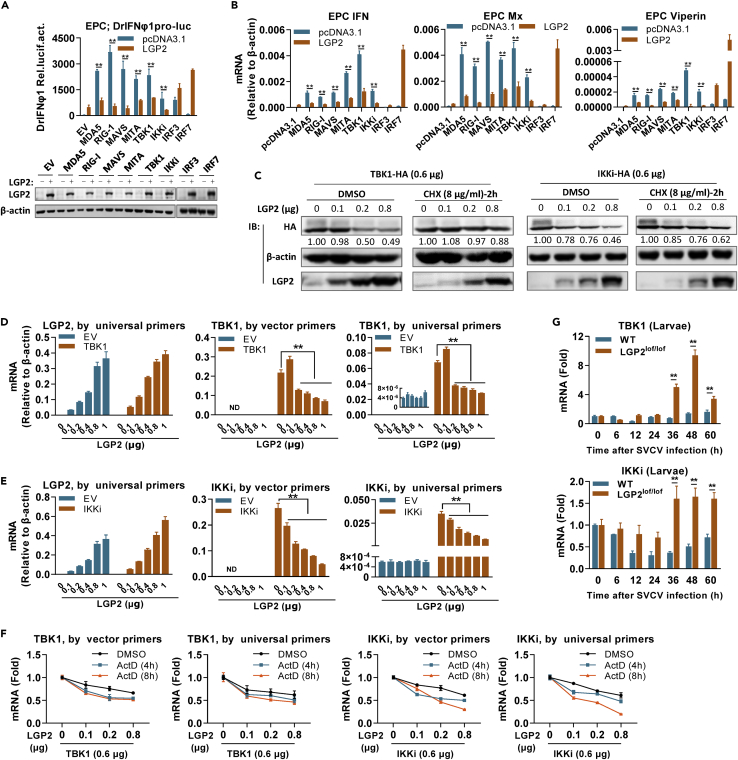
LGP2 negatively regulates IFN response by attenuating tbk1 and ikki mRNA levels
(A and B) LGP2 inhibited fish IFN promoter activation (A) and ifn expression (B) induced by RLR signaling molecules upstream of IRF3/7. EPC cells seeded in 24-well plates were transfected with DrIFNφ1pro-luc, LGP2, and each of the indicated RLR signaling molecules (200 ng each) for 48 h, followed by luciferase assays (A) or by RT-PCR detection of cellular ifn, mx, and viperin mRNA (B). Western blots in (A) showed the expression of LGP2 protein in (A and B) by western blots using anti-LGP2 Ab.
(C) LGP2-mediated protein reduction of TBK1 and IKKi was abolished by CHX. EPC cells seeded in 6-well plates overnight were transfected for 24 h with TBK1 or IKKi (0.6 μg each), together with LGP2 at increasing doses, followed by addition of CHX (8 μg/mL) or DMSO as control. Another 2 h later, cells were collected for western blotting analysis of TBK1 and IKKi by anti-HA Ab and LGP2 by anti-LGP2 Ab. The numbers show the densitometric quantification of TBK1 or IKKi protein expression normalized to β-actin.
(D and E) LGP2 attenuated mRNA levels of the transfected TBK1 (D) and IKKi (E). CO cells seeded in 6-well plates were transfected with LGP2 at increasing doses (0, 0.1, 0.2, 0.4, 0.8, 1 μg), together with TBK1 (300 ng, D) or IKKi (300 ng, E) for 48 h, followed by RT-PCR detection of lgp2, tbk1, and ikki mRNA, respectively. Universal primers were designed against the ORF sequences of tbk1 and ikki for detection of mRNA from cellular genes and the transfected plasmids together, and vector primers only detected mRNA from the transfected plasmids because a forward primer was designed against vector sequences.
(F) LGP2-mediated reduction of tbk1 and ikki mRNA levels was not impaired by ActD. EPC cells seeded in 6-well plates overnight were transfected for 24 h as in (C), followed by addition of ActD (1 μg/mL) or DMSO as control. At 48 h after ActD addition, tbk1 and ikki transcripts were detected by universal primers and vector primers.
(G) The transcript levels of tbk1 and ikki were enhanced in lgp2lof/lof zebrafish larvae during the later period of SVCV infection, in contrast to a nearly constant transcript level in WT larvae. The samples were the same as in Figure 2A. Data were shown as mean ± SD (N = 3). P values were calculated using Student’s t test. ∗∗p < 0.01. See also Figure S5 and Table S1.

