
Image
|
Figure Caption
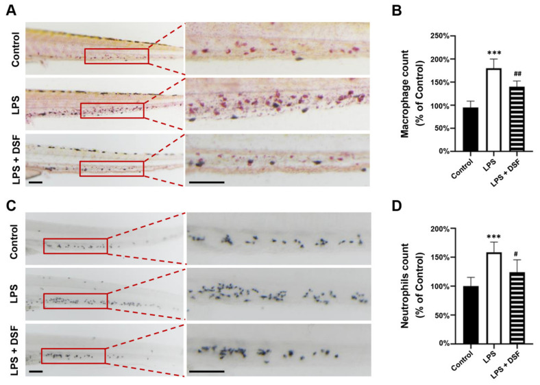
Fig. 3
DSF prevented LPS-induced accumulation of macrophages and neutrophils in zebrafish embryos. (A) The macrophages were labeled with Neutral red staining. (B) Quantitative analysis of macrophages in the same area (red boxes) in A. (C) The neutrophils were labeled with Sudan Black B staining. (D) Quantitative analysis of neutrophils in the same area (red boxes) in C. Data are represented as mean ± S.D. *** p < 0.001 vs. control group; # p < 0.05, ## p < 0.01 vs. LPS group. Scale bar, 100 μm.
Acknowledgments
This image is the copyrighted work of the attributed author or publisher, and
ZFIN has permission only to display this image to its users.
Additional permissions should be obtained from the applicable author or publisher of the image.
Full text @ Int. J. Mol. Sci.