Fig. 3
Fig. 3
Reduced cell proliferation and differentiation of CNCCs in mynn−/− embryos
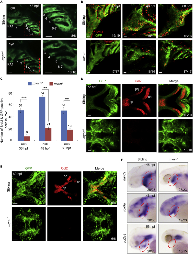
(A) Live imaging of the pharyngeal region of mynn−/− embryos in Tg(fli1:EGFP) background. The pharyngeal arches were numbered. The boxed areas in the left pannel were enlarged in the right panels. PA: pharyngeal arch. Scale bars, 20 μm.
(B and C) BrdU incorporation experiments showed reduced proliferating CNCCs in mynn−/− mutants. BrdU-treated mynn−/− mutants with fli1:EGFP expression were harvested at indicated stages and stained with anti-BrdU (red) and anti-GFP (green) antibodies. The pharyngeal regions were observed by confocal microscopy (B). Scale bars, 20 μm. The number of BrdU+ and GFP+ cells in the second pharyngeal arch was calculated from six embryos (C). Error bars indicate ±S.D. ∗∗, p < 0.01; ∗∗∗, p < 0.001 (by Student’s t test).
(D and E) Detection of Col2 proteins in the pharyngeal arches. Wild-type and mynn−/− embryos in Tg(fli1:EGFP) background at 60 (E) or 72 (D) hpf were co-immunostained with anti-GFP (green) and anti-Col2 (red) antibodies. The palatoquadrate (pq), ethmoid plate (ep), and ceratohyal (ch) cartilages were shown with anterior to the left. Scale bars, in panel D, 20 μm; in panel E, 50 μm.
(F) Expression pattern of hand2, sox9a, and col2a1 in mynn−/− embryos and their siblings at indicated stages detected by in situ hybridization.

