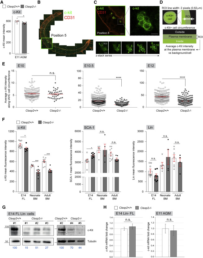
Fig. 5 Figure 5. Progressive loss of c-Kit at the plasma membrane of Clasp2−/− cells throughout development (A) MFI of c-Kit measured by flow cytometry in E11 Clasp2+/+ and Clasp2−/− AGMs (9 Clasp2+/+, 7 Clasp2−/−, n = 7). (B) Tile-scale image reconstruction of a whole E10.5 Clasp2+/+ embryo stained with c-Kit (green) and CD31 (red) antibodies. (C) Enlarged image of position 5 shown in (B), with a dashed box outlining the inset, which is shown enlarged on the right and reveals c-Kit fluorescence (green) in an IAHC. Bottom panels: images of this IAHC in various focal planes to draw regions of interest (ROIs) at the maximal fluorescence intensity along the circumference of IAHC cells through a z stack series. (D) Illustration of a IAHC cell with the ROI (2 pixels in width) to measure the average c-Kit fluorescence intensity. (E) Average MFI of c-Kit along the plasma membrane circumference of IAHC cells in the aortae of E10, E10.5, and E12 Clasp2+/+ and Clasp2−/− embryos after whole-mount immunostaining with c-Kit antibody (E10: 1 Clasp2+/+, 2 Clasp2−/−; E10.5: 1 Clasp2+/+, 1 Clasp2−/−; E12: 1 Clasp2+/+, 1 Clasp2−/−). (F) MFI of c-Kit, SCA-1, and Lin markers, measured by flow cytometry in E14 FL and neonate and adult BM cells isolated from Clasp2+/+ and Clasp2−/− embryos and mice (E14 FLs [8 Clasp2+/+, 6 Clasp2−/− embryos], n = 3; P8 BM [6 Clasp2+/+, 8 Clasp2−/−], n = 6; adult BM [10 Clasp2+/+, 6 Clasp2−/−], n = 7). (G) WBs for c-Kit and tubulin (loading control) on lysates of E14 Clasp2+/+ and Clasp2−/− Lin− FL cells (2 Clasp2+/+, 5 Clasp2−/−, n = 2). c-Kit intensity is indicated in blue below the gel images; the Clasp2+/+ culture band was set at 100. (H) qRT-PCR for c-Kit on Lin− FL cells and total AGM cells from Clasp2+/+ and Clasp2−/− embryos isolated at E14 and E11, respectively. Error bars: mean ± SEM (A, E, and F), mean ± SD (H). ∗∗∗∗p < 0.0001, ∗∗∗p < 0.001, ∗p < 0.05, Mann-Whitney U test (A, E, and F), unpaired t test (H). Scale bars, 100 μm (B and C) and 10 μm (C, close up).
Image
Figure Caption
Acknowledgments
This image is the copyrighted work of the attributed author or publisher, and
ZFIN has permission only to display this image to its users.
Additional permissions should be obtained from the applicable author or publisher of the image.
Full text @ Cell Rep.

