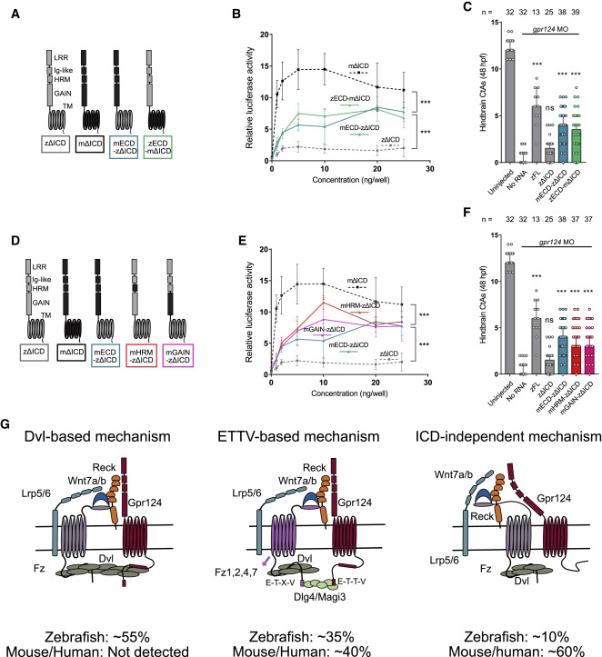
Fig. 7 Figure 7. Functional comparison of mouse and zebrafish Gpr124 extracellular and transmembrane domains (A and D) Schematics of the investigated mouse and zebrafish Gpr124 chimeras. (B and E) Luciferase dose-response assays in HEK293 STF cells co-transfected with Wnt7a, Fz1, Reck, and the indicated Gpr124 chimeras. Data are normalized to ELISA data, n = 3. (C and F) Quantification of hindbrain CtAs in 48 hpf gpr124 morphant embryos injected at the one-cell stage with 100 pg of the indicated mRNAs. (G) Model for the various modalities of Gpr124/Reck-dependent Wnt7a/b signaling and the importance of their relative contribution in different species. Related to Figures S5 and S6.
Image
Figure Caption
Figure Data
Acknowledgments
This image is the copyrighted work of the attributed author or publisher, and
ZFIN has permission only to display this image to its users.
Additional permissions should be obtained from the applicable author or publisher of the image.
Full text @ Cell Rep.

