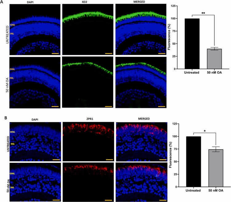
Fig. 8 Fig. 8. (A) Detection of rod cells visualization by immunostaining with anti-rhodopsin 4D2 antibody in zebrafish embryos treated with or without 50 nM OA from 24 to 120 hpf. (B) Cone cells were detected by immunostaining with ZPR-1 antibody in retinal sections of zebrafish embryos treated with or without 50 nM OA from 24 to 120hpf. Embryos exposed to OA illustrated a reduction in fluorescence intensity. Five images were taken from untreated and treated zebrafish retinal sections and quantified using Image J software. Data was presented as mean ± SEM (n = 5). * p < 0.05; * * p < 0.01. Scale bar, 20 µm.
Image
Figure Caption
Figure Data
Acknowledgments
This image is the copyrighted work of the attributed author or publisher, and
ZFIN has permission only to display this image to its users.
Additional permissions should be obtained from the applicable author or publisher of the image.
Full text @ Toxicology

