IMAGE

FIGURE 3

ID
ZDB-IMAGE-220424-3
Genes
Source
Figures for Ouyang et al., 2022
Image
Figure Caption

FIGURE 3

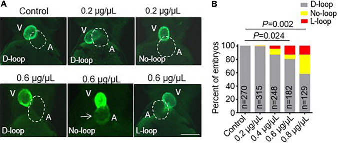
CPAAHTs exposure induced ectopic expression of vmhc gene in the atrium and caused cardiac-looping defects to dose-dependently. (A,B) Representative images of heart-looping morphology (A) and quantification analysis (B) of the percentage of the vmhcKI–EGFP KI embryos with corresponding cardiac-looping structure after exposure to different concentrations of CPAAHTs compared to untreated control at 48 hpf. The arrow points to the ectopic expression of the EGFP signal in the atrium. The dotted line outlines the shape of the atrium. V, ventricle. A, atrium. N = 129–315, chi-squared test.

Figure Data
Acknowledgments
This image is the copyrighted work of the attributed author or publisher, and ZFIN has permission only to display this image to its users. Additional permissions should be obtained from the applicable author or publisher of the image. Full text @ Front Cardiovasc Med