IMAGE

Fig. 2.

ID
ZDB-IMAGE-220331-11
Source
Figures for Kugler et al., 2022
Image
Figure Caption

Fig. 2.

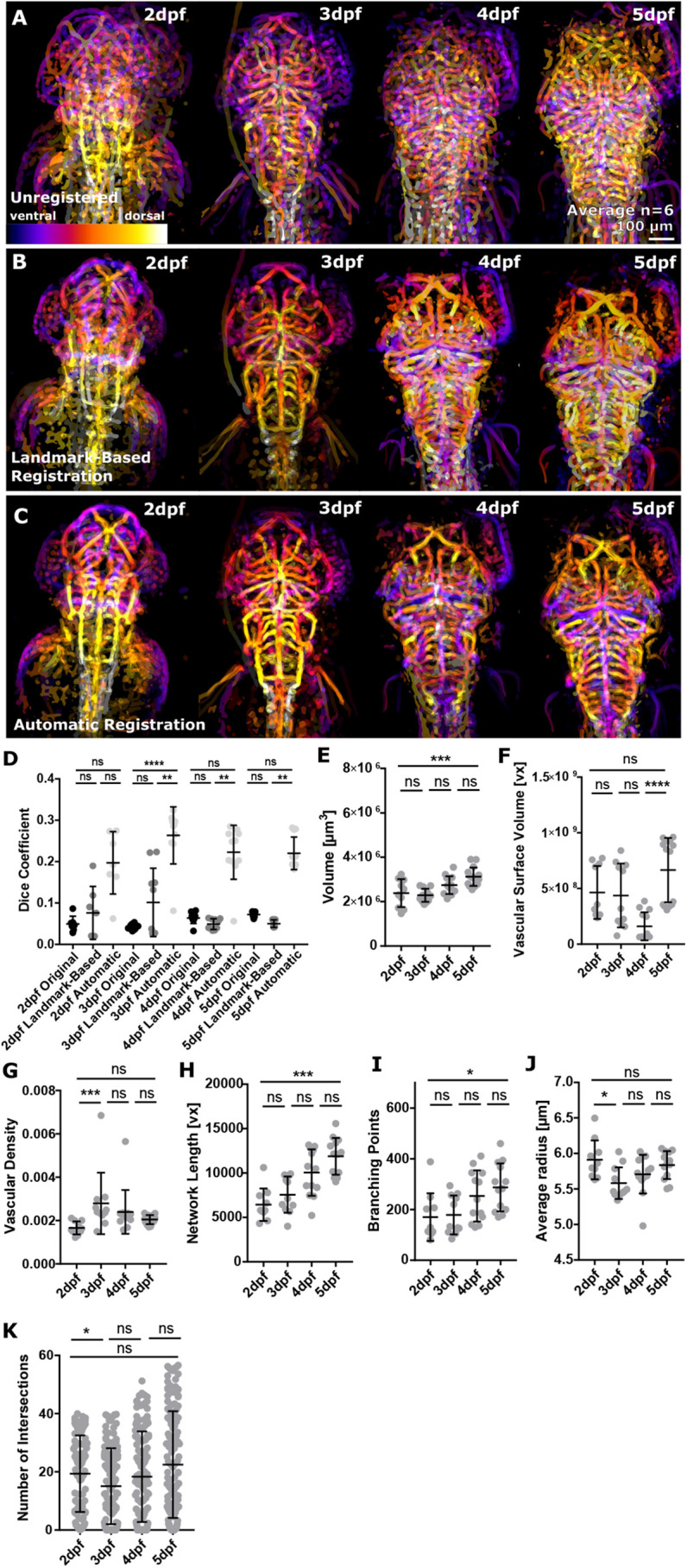
Application of ZVQ to embryos from 2 to 5 dpf to analyse vascular growth. (A) Depth-coded MIP showing regions of overlap (purple, ventral; white, dorsal) of six fish before registration from 2 to 5 dpf. (B) Depth-coded MIP showing regions of overlap (purple, ventral; white, dorsal) of six fish after manual registration from 2 to 5 dpf. (C) Depth-coded MIP showing regions of overlap (purple, ventral; white, dorsal) of six fish after automatic registration from 2 to 5 dpf. (D) Dice coefficient between template and moving image was increased after application of rigid registration using both anatomical landmark-based and automatic rigid registration from 2 to 5 dpf [2 dpf, n=7; 3 dpf, n=10; 4 dpf, n=10; 5 dpf, n=10; two experimental repeats; Kruskal–Wallis test; ns (P>0.5), **P=0.01-0.001, ****P<0.0001; data are mean±s.d.]. (E) Vascular volume was statistically significantly increased from 2 to 5 dpf (P=0.0008; 2 dpf, n=10; 3 dpf, n=12; 4 dpf, n=13; 5 dpf, n=15; one-way ANOVA; data are mean±s.d.). (F) Vascular surface was not statistically significantly increased from 2 to 5 dpf (P=0.4885; 2 dpf, n=10; 3 dpf, n=11; 4 dpf, n=13; 5 dpf, n=14; two experimental repeats; Kruskal–Wallis test; data are mean±s.d.). (G) Vascular density was not statistically significantly increased from 2 to 5 dpf (P=0.2041; 2 dpf, n=10; 3 dpf, n=11; 4 dpf, n=13; 5 dpf, n=14; two experimental repeats; Kruskal–Wallis test; data are mean±s.d.). (H) Network length was statistically significantly increased from 2 to 5 dpf (P=0.0001; 2 dpf, n=10; 3 dpf, n=12; 4 dpf, n=13; 5 dpf, n=15; Kruskal–Wallis test; mean±s.d.). (I) Branching points were statistically significantly increased from 2 to 5 dpf (P=0.0082; Kruskal–Wallis test; data are mean±s.d.). (J) Average vessel radius was not statistically significantly changed from 2 to 5 dpf (*P>0.9999; Kruskal–Wallis test; data are mean±s.d.). (K) Sholl analysis was conducted to assess vascular complexity, showing no significant increase from 2 to 5 dpf (2-3 dpf, *P=0.0340; 3-4 dpf, P=0.6825; 4-5 dpf P=0.2000; 2-5 dpf, n=5; Kruskal–Wallis test; data are mean±s.d.).

Acknowledgments
This image is the copyrighted work of the attributed author or publisher, and ZFIN has permission only to display this image to its users. Additional permissions should be obtained from the applicable author or publisher of the image. Full text @ Development