Figure 1
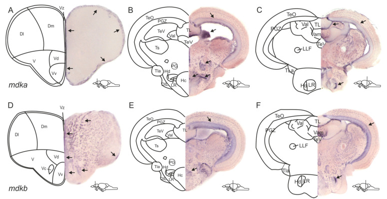
Expression patterns of the two paralogues mdka and mdkb differ in the adult zebrafish brain. (A–F) In situ hybridization (ISH) against mdka (A–C) and mdkb (D–F) on cross-sections through the zebrafish adult brain. mdka staining is visible along the ventricular layers of the dorsal and ventral telencephalic areas (A) and in the periventricular grey zone of the optic tectum and the lateral recess of the periventricular hypothalamus (B,C). In the diencephalon, mdka expression is additionally observed in the valvula cerebelli, the preglomerular nucleus, and the periventricular hypothalamus (B). mdkb expression is also detected in and outside of the ventricular layers of the telencephalon and spreads into the nuclei of the ventral telencephalon (D). In the diencephalon, it is also visible in the periventricular grey zone and the lateral recess (E,F). Abbreviations: D: dorsal telencephalic area; Dl: lateral zone of D; DIL: diffuse nuclei of the inferior lobe; Dm: medial zone of D; Hc: caudal zone of the periventricular hypothalamus; Hd: dorsal zone of the periventricular hypothalamus; LLF: lateral longitudinal fascicle; LR: lateral recess of the diencephalic ventricle; PG: preglomerular nucleus; PGZ: periventricular grey zone of TeO; TeO: optic tectum; TeV: telencephalic ventricle; TL: torus longitudinalis; TLa: torus lateralis; Ts: torus semicularis; V: ventral telencephalic area; Val: lateral division of the valvula cerebelli; Vam: medial division of the valvula cerebelli; Vc: central nucleus of V; Vd: dorsal nucleus of V; Vv: ventral nucleus of V; Vz: ventricular zone of the telencephalon.

