Figure 1
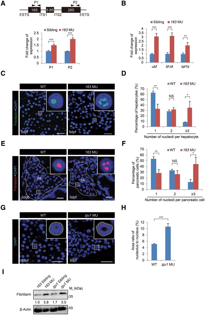
Loss-of-function of Bms1l upregulates the pre-rRNA transcription and increases the volume of nucleoli in hepatocytes. (A) qPCR analysis of total rRNA transcripts in 5dpf-old bms1lsq163/sq163 mutant and its siblings (the pool of bms1+/+ and bms1sq163/+, used thereafter) using two pairs of primers derived from 18S (P1) and 28S (P2) showing an upregulation of rRNA expression in bms1lsq163/sq163 mutant. Upper panel, a diagram showing the genomic structure of the zebrafish rDNA gene and the positions of P1 and P2. (B) qPCR analysis showing the upregulation of ubf, tif-IA, and taf1b expression in 5dpf-old bms1lsq163/sq163 mutant compared with the siblings. The qPCR values were normalized against GAPDH and expressed as fold change of expression. The values plotted represent mean ± SEM. **P < 0.01, ***P < 0.001. (C–F) Fibrillarin immunostaining and DAPI staining (green in C and red in E) showing the significant increase in the number of nucleoli in bms1lsq163/sq163 hepatocytes (D, three WT embryos, 1397 cells examined; three bms1lsq163/sq163 embryos, 1274 cells examined) and pancreatic cells (F, three WT embryos, 1015 cells examined; three bms1lsq163/sq163 embryos, 463 cells examined) when compared with WT, respectively, at 5dpf. The pancreatic region (pa) was outlined by a dashed line. Scale bar, 20 μm (C) and 10 μm (E). (G and H) DAPI staining (G) showing the significant increase in the size of nucleolus in bms1lzju1/zju1 hepatocytes compared with WT at 5dpf (H, three WT embryos, 112 cells examined; four bms1lzju1/zju1 embryos, 119 cells examined). Scale bar, 20 μm. (I) Western blotting showing the upregulation of Fibrillarin protein levels in 5dpf-old bms1lsq163/sq163 and bms1lzju1/zju1 mutants compared with their siblings. β-Actin: loading control. Insets in C, E, and G showing higher magnification of a representative nucleus (boxed). The values in D, F, and H plotted represent mean ± SEM. *P < 0.05, **P < 0.01, ***P < 0.001; NS, no significance.

